9 Jul 2018
James Warland discusses the identification and management of comorbidities in cats with this condition.
James Warland
Job Title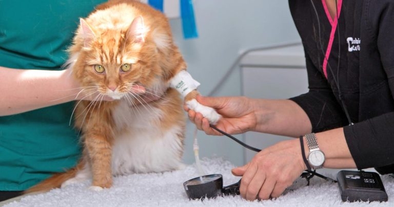
Feline hyperthyroidism is a common condition in general practice and usually straightforward to diagnose. However, management can be challenging due to variable responses, owner preferences, side effects and comorbidities, necessitating an individualised approach to each case and careful explanation of options to owners.
Feline hyperthyroidism is a common condition in general practice and usually straightforward to diagnose. However, management can be challenging due to variable responses, owner preferences, side effects and comorbidities, necessitating an individualised approach to each case and careful explanation of options to owners.
Hyperthyroidism is the most common endocrinopathy in cats, with up to 10% of geriatric cats developing the condition in their lives1.
Most clinicians are aware of the high probability of this condition in an older cat showing typical clinical signs, such as increased appetite, weight loss, polyuria/polydipsia, gastrointestinal (GI) signs and behavioural changes.
With increasing clinical awareness, hyperthyroidism is commonly diagnosed with routine screening or in cats showing very mild clinical signs. Given this increased recognition, two-thirds of hyperthyroid cats reported in a large cohort were of ideal body condition or overweight, although the majority had shown a reduction in weight2.
A few cats will show “apathetic“ hyperthyroidism, with poor appetite and lethargy. Hyperthyroidism, or its treatment, can cause wide-ranging clinical effects, such as on the cardiovascular and renal systems, making management more challenging. As well as direct effects, geriatric cats will frequently suffer from comorbidities that can further complicate the management of hyperthyroidism.
Comorbidities can either be a direct consequence of the hyperthyroid state or incidental – reflecting the older age of these patients. Cardiac disease – particularly tachycardia, arrhythmia and hypertrophy – are well-recognised consequences of hyperthyroidism. Chronic kidney disease (CKD) is frequently recognised in cats with hyperthyroidism, but is also common in the non-hyperthyroid geriatric population.
It is unclear to what extent CKD and hyperthyroidism represent independent, comorbid conditions, and whether hyperthyroidism drives significant renal injury. The impact of treatment (or lack of treatment) of hyperthyroidism on renal disease progression is an important consideration when managing cases.
Most studies considering comorbidities in hyperthyroid cats are from referral centres usually with a focus on radioactive iodine treatment (RIT). This must be considered when applying these results to the majority of cases treated in general practice.
A UK report3 found around 35% of cases to have hypertension and a similar number to have renal disease. Interestingly, these comorbidities were not related to the severity of the hyperthyroidism. Cardiac disease was seen with increasing frequency (37% versus 71% in mild versus severe hyperthyroidism) and consequence (0% versus 16% dyspnoeic), with increasing hyperthyroidism severity.
The value in further imaging (radiography, ultrasound) screening for comorbidities is debated. Given the expense and long hospitalisation involved in treatment, radioiodine patients are frequently screened to rule out concurrent disease before treatment. Puig et al4 found 18% (17 of 94) cases presented for RIT, but without overt comorbidities, had concurrent disease directly leading to altered clinical management (not pursuing RIT). The most commonly identified conditions were chronic enteropathy (4 of 17) and alimentary lymphoma (5 of 17). In the same study, 10% of cats were found to have asymptomatic urinary tract infections when culture was performed, although this merely delayed RIT4.
In contrast, a study evaluating abdominal ultrasound5 found, while 36% (193 of 534) had abnormal findings, only 2.2% (12 of 534) were not treated as a result. Neoplasia was infrequently found (2.4%) and was not always a barrier to RIT.
The author takes a practical view that screening for concurrent disease should always be considered, but with greater emphasis placed on patients with concerning clinical signs or physical examination findings (for example, GI signs, heart murmur or abnormal abdominal palpation) and on candidates for treatment that is invasive, expensive or likely to impact on quality of life (for example, thyroidectomy and RIT).
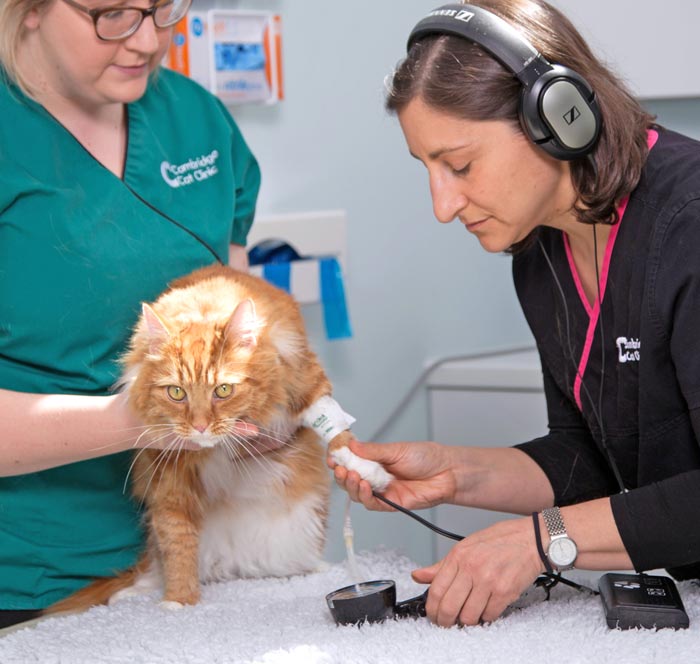
All cats diagnosed with hyperthyroidism should, as a minimum, have a full physical examination, blood pressure assessment (Figure 1), haematology, biochemistry and urinalysis (ideally including culture and protein:creatinine ratio). Further imaging should be performed on a case-by-case basis.
While the prevalence of renal insufficiency appears to be similar between hyperthyroid cats and age-matched controls, it is unclear to what extent hyperthyroidism is damaging to kidney function versus being concurrent diseases in ageing cats. A review of renal function in feline hyperthyroidism discusses this in detail6.
Hyperthyroidism is recognised to cause activation of the renin-angiotensin-aldosterone system, leading to increased cardiac output, decreased vascular resistance and increased blood volume. These factors, along with other mechanisms, lead to increased renal blood flow and glomerular filtration rate (GFR). The increased GFR, which can reduce serum creatinine to normal concentrations, is said to “mask“ CKD. Decreased muscle mass also contributes to this effect, given associated reductions in creatinine.
Proteinuria is a common finding in hyperthyroid cats, although the mechanism is not well understood. Proteinuria is recognised to be a risk factor for the development and progression of CKD. Urinary markers of tubular injury, such as N-acetyl-β-D-glucosaminidase, are elevated in hyperthyroidism and decrease after treatment, suggesting a deleterious effect of the hyperthyroid state on the kidneys7.
It is well recognised many non-azotaemic, hyperthyroid cats develop azotaemia following restoration of euthyroid status. Around 25% of well-controlled euthyroid cats will develop azotaemia over the eight months after initiating therapy8. Despite the worsening of renal parameters, the return to euthyroidism is considered to be beneficial to renal function.
Recognising renal insufficiency is important, as cats with documented azotaemia at diagnosis have shorter survival times than those with normal renal parameters8. However, the same study found no evidence that, after restoring euthyroid status, the subsequent development of azotaemia reduces life expectancy9. Using the International Renal Interest Society (IRIS) guidelines, most cats will show a predictable increase of up to one IRIS stage, after hyperthyroidism treatment, which then remains stable10,11. Therefore, the concern regarding the development of overt CKD after treatment has likely been overstated. Given the detrimental effect of hyperthyroidism on renal health, cats with CKD should always have their hyperthyroidism managed.
Iatrogenic hypothyroidism has a negative impact on survival in cats that develop azotaemia, although not in cats without renal insufficiency9. Restoration of euthyroidism in cats with iatrogenic hypothyroidism is associated with an improvement in renal function12, and a study found azotaemic, hypothyroid cats supplemented with levothyroxine lived significantly longer than those left untreated13. Taken together, it is clear iatrogenic hypothyroidism is detrimental in cats with CKD and clinicians should be alert to the development of this complication, particularly where renal disease is present. Further discussion of iatrogenic hypothyroidism is included in part two of this article.
Significant research has focused on predicting which cats will show renal insufficiency after treatment. Cats with higher pre-treatment creatinine are at higher risk of this increasing to the point of being classified as azotaemia8. Despite being routinely touted and logical parameters to consider, neither proteinuria nor low urine-specific gravity (USG) are convincingly associated with the development of azotaemia after treatment in several studies8,14, although low USG has been associated with post-treatment azotaemia in one publication15. However, proteinuria has been associated with an increased all-cause mortality8.
Symmetric dimethylarginine (SDMA) greater than 14μg/dL, has been shown to be a specific (98%) marker for post-treatment renal insufficiency (in cats non-azotaemic at diagnosis), and cats with high SDMA are highly likely to develop azotaemia15. Sensitivity was still low (33%), meaning most cats were not identified prior to treatment. The author recommends screening cats after diagnosis with a full urinalysis, including for proteinuria and silent urinary tract infection, to provide a full clinical picture and provide prognostic information, despite the difficulty in predicting azotaemia with this information.
Similarly, although SDMA may not change the clinical management, it may be valuable to alerting owners to the presence of CKD before undertaking hyperthyroidism treatment.
Practically, in many cases, the only way to reliably identify occult renal disease is to treat the hyperthyroidism. For this reason, it is frequently recommended to use medical therapy (which can be reduced/stopped) prior to using permanent solutions (surgery/RIT).
This approach is probably unnecessary given the recognised need for hyperthyroidism treatment even in CKD, rises in creatinine being rarely more than one IRIS stage, and survival not being affected by the development of azotaemia. Therefore, treatment with RIT will rarely be contraindicated by the unmasking of renal disease in these cats. Many owners, however, will want as full a picture as possible of renal health before undertaking permanent treatment, and this medical treatment trial can be helpful.
It may be advisable to begin medical treatment relatively gently, in an attempt to avoid a precipitous drop in GFR and hypothyroidism, especially in cases with evidence of significant renal disease. The hyperthyroid state is considered to be deleterious to renal health; therefore, previous recommendations of not treating or “under-treating“ hyperthyroidism in cats with renal disease – with the intention of increasing GFR – are not recommended. Renal parameters should always be monitored alongside T4 when assessing response to treatment, and should be regularly checked for at least six months after stabilising the patient.
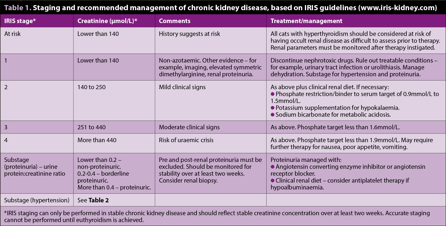
The author would recommend renal disease is classified and treated according to the IRIS guidelines (www.iris-kidney.com; Table 1), with staging of CKD performed once stability has been achieved.
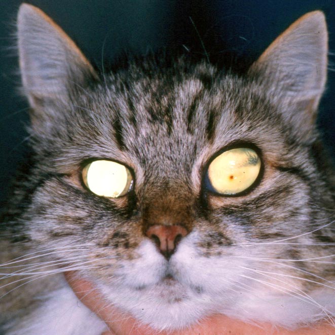
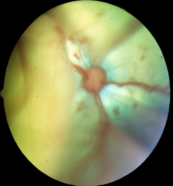
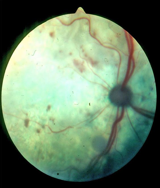
The cause of hypertension in hyperthyroid cats is unclear, and likely to be multifactorial. The consequences of high blood pressure on “target organs“ can be devastating, including retinal haemorrhage/detachment (and blindness; Figure 2), renal injury, cardiac hypertrophy and brain injury. Hypertension in hyperthyroidism is associated with higher mortality8, and while definitive evidence that treatment improves survival is lacking, management is warranted, given the potential consequences.
Elevated blood pressure is reported in 10% to 25% of cats at diagnosis16 with a further 25% developing hypertension after treatment17. Cats must be monitored for hypertension both before and after hyperthyroidism treatment. While CKD is a risk factor for hypertension, 61% of the cats that developed hypertension after treatment did not develop concurrent azotaemia. Physical examination of cats at risk of hypertension should include fundoscopy.
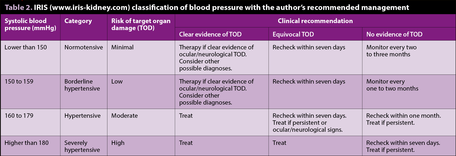
Guidelines for the management of hypertension in cats have been published16 (Table 2), including practical guidance on achieving accurate measurements, which are notoriously susceptible to the “white coat effect“ of stress-induced elevation. Cats should have their blood pressure measured – ideally with a Doppler system, as this is the most accurate and well-validated system. Systolic blood pressure (SBP) measurement is the most accurately measured and widely reported, so is considered more valuable than mean or diastolic pressures.
Treatment of hypertension should be instigated in cases where SBP is greater than 150mmHg to 160mmHg on two occasions, if target organ damage (TOD) is suspected (no need to confirm persistence if clear evidence of TOD), or if persistently (two separate measurements, over one week) greater than 180mmHg and a “white coat effect“ is not suspected, even in the absence of TOD. If persistently greater than 160mmHg on several measurements longer than one month, the author would also treat the hypertension in the absence of TOD. During stabilisation, blood pressure should be checked at least weekly. Once stable, blood pressure should be monitored every three to six months. Target SBP should be 110mmHg to 150mmHg.
Treatment recommendations for hypertension are the same, whether hyperthyroid or euthyroid. Recommended initial therapy is amlodipine (0.625mg/cat/day PO or 1.25mg/cat/day if SBP is greater than 200mmHg). Most cats will respond to therapy with typical SBP reductions of 30mmHg to 70mmHg16. The dose can be adjusted up as necessary. Up to 2.5mg/cat/day can be given, although the author would usually add a second agent if inadequate control is achieved with 1.25mg/kg.
Angiotensin-converting enzyme inhibitors (for example, benazepril 0.5mg/kg/day PO to 1mg/kg/day PO) or angiotensin receptor blockers (for example, telmisartan 1mg/kg/day PO) can be used in addition to amlodipine. Care must be taken to avoid hypotension. Some cases with severe hypertension and TOD may require hospitalisation and emergency therapy to bring down SBP within hours. Initial therapy can be with amlodipine as aforementioned, but parenteral treatment (for example, hydralazine) should be considered.
The importance of rechecking blood pressure after treatment of hyperthyroidism cannot be overstated. Many cats will be normotensive at diagnosis and develop hypertension after treatment – this must be recognised and treated to ensure optimum outcomes are achieved. Alternative causes of hypertension, such as primary hyperaldosteronism, should also be considered.
Liver enzyme activity is frequently elevated in cats with hyperthyroidism and has been reported in several studies, with greater than 80% having an increased alanine aminotransferase activity1. The author would consider further investigation of liver disease if the degree of elevation was disproportionate to the hyperthyroidism, or other clinical signs or physical examination findings were consistent with hepatic disease.
Cardiomyopathy with ventricular hypertrophy, arrhythmia and thromboembolic disease is seen with cats with hyperthyroidism. Overt congestive heart failure appears to be uncommon, but likely increases with severity and chronicity of hyperthyroidism. Asymptomatic cases rarely require additional therapy (beyond treatment of hyperthyroidism), although consideration should be given to antiplatelet therapy if risk factors for thromboembolism are identified (for example, left atrial spontaneous echogenic contrast).
Interpretation of cardiac investigations, including echocardiography and N-terminal pro-B-type natriuretic peptide measurement, can be challenging given significant overlap between primary cardiac disease and changes secondary to hyperthyroidism18. Treatment for congestive heart failure thought to be secondary to hyperthyroidism should be as for primary cardiac disease.
When assessing cardiac disease in cats with hyperthyroidism, the focus should be on whether congestive cardiac failure is present and requiring treatment, as a definitive cardiac diagnosis is unlikely to be achievable until they are euthyroid and stable for several months. Echocardiographic changes due to hyperthyroidism are usually reversible during the months after euthyroidism is achieved18. The presence of cardiac disease or failure should not affect the treatment of hyperthyroidism per se, but may affect the suitability of the patient for certain treatments, such as surgery (and general anaesthesia).
The identification and management of comorbidities are vital to ensuring optimal management of hyperthyroidism. Renal disease and hypertension are frequently encountered, but clinicians must remember to monitor for these after treatment of hyperthyroidism, as they may only be revealed after treatment is instigated. Importantly, renal disease should not be seen as a reason to under-treat the thyroid disease, as this has further detrimental effects on renal function. Iatrogenic hypothyroidism must be avoided in these cases.
Part two of this article will discuss different treatment options to help make the best choice for individual patients.
The author would like to thank Mini Wright and Tim Williams for their critical appraisal of the article and suggested improvements. The author is also very grateful to David Williams, Yordan Fernandez and Dr Wright for the images used.