10 Jul 2017
Floor Driessen discusses the case of a seven-year-old male, neutered greyhound in the latest in the Case Notes series.
Floor Driessen
Job Title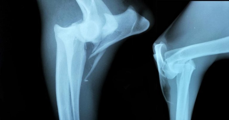
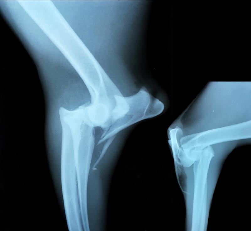
Figure 1. Lateral (left) and craniocaudal views of the right elbow.
A seven-year-old male, neutered greyhound is presented with acute onset non-weightbearing thoracic limb lameness following a fall down a muddy bank.
In clear discomfort, swelling and bruising is noted around the elbow, with asynchronous movement in the distal limb. On clinical examination, crepitus associated with manipulation of the elbow is noted. Radiographs of the elbow are presented in Figure 1.
What is this fracture called and how would it be classified? Are further investigations indicated, and how may this be treated?
The radiographs show comminution of the proximal part of the ulna, extending to the olecranon. Due to the pull of the triceps, the proximal ulnar fragment and anconeal process have been displaced proximally.
Concomitant cranial luxation of the radial head is present. The nature of the injury suggests damage to the medial collateral ligament has also occurred.
The specific combination of fracture of the proximal third of the ulna and concurrent luxation of the radial head is known as a Monteggia fracture – named after Italian surgeon Giovanni Battista Monteggia, who described the injury in 1814.
While a history of known trauma in this particular case exists, the magnitude of the trauma did not appear
to correlate with the severity of the injury. Additionally, the age and breed of the dog raised suspicion for the possibility of a pathological fracture.
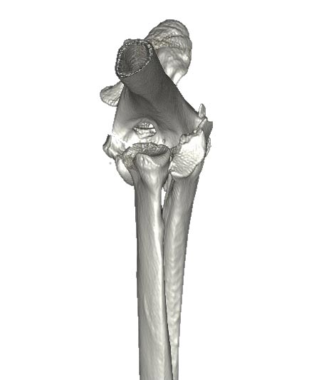
To further characterise the injury, a CT scan was performed (Figure 2). It found no evidence of bone lysis or periosteal reaction at the fracture site, so a pathological fracture due to malignancy was ruled out.
The scan showed a fragment of bone opacity medial to the medial epicondyle of the humerus and, in combination with physical examination, was suspected to represent partial avulsion of the medial collateral ligament.
Monteggia fractures are defined as an ulnar fracture with radial head luxation. It is uncommon in dogs and cats, and believed to occur most commonly if a force has been applied on the caudal part of the ulna during weightbearing.
Although further subdivisions exist, four classifications are commonly used in veterinary orthopaedics:
Concurrent damage to ligaments can be seen. Stretching and tearing of the annular ligament can occur and, when the radius and ulna are separated, damage to the interosseous ligament may be seen.
In this case, increased laxity without complete rupture of the medial collateral ligament was noted, suggesting a higher grade sprain.
As with all articular fractures, the goal of treatment is precise restoration of the articular surface. In Monteggia fractures, restoration of humeroradial joint congruity is also required. As such, rigid internal fixation is preferred. Collateral and interosseous ligament reconstruction may also be required.
With accurate reconstruction, such fractures carry a fair to good prognosis. If comminution is severe, or access to expertise limited, such fractures may not be reconstructable; amputation remains a treatment option in such cases.