24 Oct 2016
Sebastien Behr looks at signs, diagnosis and treatment of masses found in the pituitary gland.
Sebastien Behr
Job Title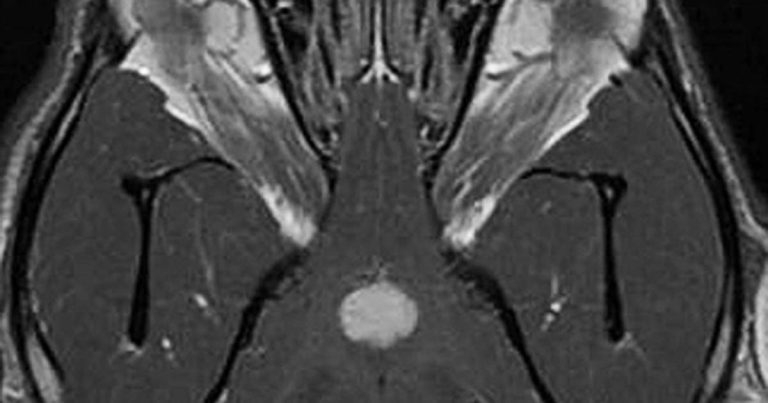
Figure 2. Dorsal 3D fast imaging employing steady-state acquisition post-contrast MRI of the brain of a dog affected by a pituitary tumour.
The pituitary gland is located in the sella turcica, within the basisphenoid bone, below the hypothalamus and in close vicinity to the optic nerve (CN II), ophthalmic branch of the trigeminal nerve (CN V), oculomotor (CN III) and trochlear nerve (CN IV).

As a result, pituitary gland tumours, leading to gland enlargement, can account for neurological signs by compression of the diencephalon and surrounding cranial nerves. They are frequent in dogs, with about 50% of canine pituitary adenomas occurring in brachycephalic breeds, but are rare in cats.
In dogs with endocrine-active pituitary tumours, the most common endocrine abnormality is hypersecretion of adrenocorticotropic hormone, resulting in Cushing’s disease. In cats, hypersecretion of growth hormone, resulting in acromegaly, can also occur. No association has occurred between endocrine test results and the size of pituitary tumours. In dogs, these tumours are usually chromophobic microadenomas (less than 1cm in diameter) that do not produce neurological signs.
Neurological signs secondary to pituitary macro-tumours are often insidious in onset and non-specific. Vague signs of central nervous dysfunction – such as lethargy, inappetence and mental dullness – were more commonly associated with pituitary macro-tumours than more specific neurological signs, such as seizure or blindness. The neurological signs can often be minimal, despite development of large pituitary tumours.
MRI study results have suggested up to 60% of dogs with pituitary-dependant macroadenoma have pituitary tumours 4mm to 12mm in diameter, without neurological signs. Considering the location of the lesion within the middle cranial fossa, the neurological signs are often symmetrical and bilateral – as, for example, bilateral proprioceptive deficits, bilateral decreased to absent menace response.
Signs of raised intracranial pressure can sometimes be noticed, such as head pressing, anisocoria and papilloedema.

A more acute and severe neurological presentation can be seen in dogs suffering from pituitary apoplexy. In such cases, an acute haemorrhage in the pituitary tumour leads to acute alteration in mental status (obtundation or stupor) and acute onset of forebrain signs (for example, decreased/absent menace response, circling and proprioceptive deficits).
This acute presentation, due to sudden change in pituitary tumour size, can alter the paired cavernous sinuses on the floor of the calvarium and lead to cavernous sinus syndrome. In such cases, neurological deficits can include external and internal ophthalmoplegia (due to lesions of CN III, IV and VI), sensory deficits of the face (due to lesions of the ophthalmic and maxillary branches of the CN V) and, more rarely, atrophy of the masticatory muscle (due to the mandibular branch of the CN V). Menace response is usually preserved, but vision can be altered due to the ophthalmoplegia.
Diagnosis of pituitary tumours relies on advance imaging, such as CT (Figure 1) or MRI of the brain (Figure 2). Small tumours are better identified on MRI than CT. It has been estimated at least 15% to 20% of dogs with pituitary-dependent macroadenoma may develop clinical signs due to a growing pituitary tumour during the first two or three years after diagnosis, such as pacing, lethargy, aimless wandering, hiding, tight circling, head pressing and seizures.
Treatment of pituitary masses/tumours can be performed by radiation therapy. When compared with untreated dogs, radiotherapy increased survival and controlled neurologic signs in dogs with pituitary masses. Mean survival time in the treated dogs was 1,405 days with one, two and three-year estimated survival of 93%, 87% and 55%, respectively (Kent et al, 2007).