21 Jan 2019
Francesco Cian presents another case in his latest Haematology Hub, this time on a mixed breed, male dog with clinical history of inappetence, anorexia and recurrent episodes of vomiting.
Francesco Cian
Job Title
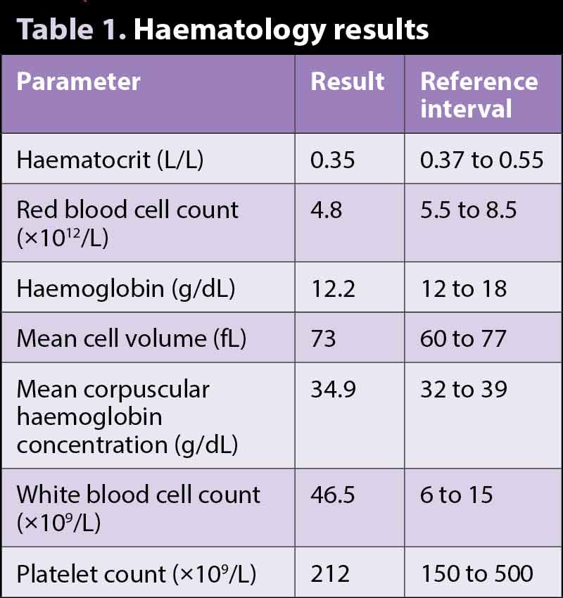
The two images (Wright-Giemsa 50×) and the data (Table 1) are from an ethylenediaminetetraacetic acid (EDTA) blood sample of an adult, mixed breed, male dog with clinical history of inappetence, anorexia and recurrent episodes of vomiting.
Based on the data and blood smear pictures, try to answer the following questions:
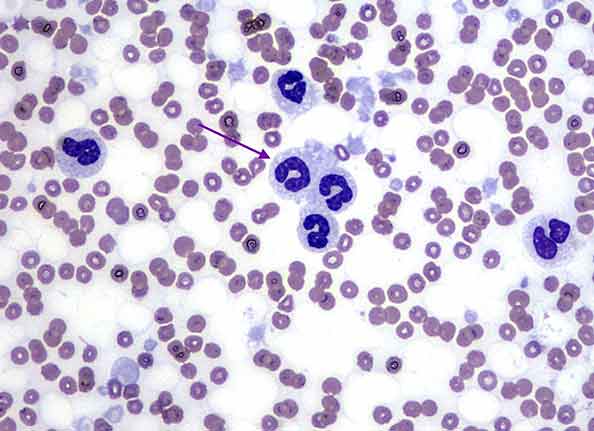
Evidence of marked leukocytosis exists, confirmed on blood smear examination. Leukocytes are mostly neutrophils and show toxic changes (red arrow), which are characterised by foamy basophilic cytoplasm and Dohle bodies. The latter are dark bluish, angular and small cytoplasmic inclusions composed of retained aggregates of rough endoplasmic reticulum.
Immature forms, called band neutrophils (purple arrow), are also present, indicating a left shift. All these changes are the result of the early release of neutrophils by the bone marrow in response to inflammation and can be recognised only by examining the blood smear under the microscope.
Monocytes (green arrow) are also increased in numbers.
Neutrophilia with left shift, toxic changes and monocytosis are overall supportive of an inflammatory leukogram. Abdominal ultrasound and pancreatic lipase immunoreactivity (PLI) measurement confirmed the presence of pancreatitis, which was the likely cause for these changes.
Evidence of persistent, mild anaemia exists, which appears normocytic normochromic, as both blood cell indices (mean cell volume and mean corpuscular haemoglobin concentration) are within reference range. This is confirmed by examination of the blood smear, which contains only rare polychromatophils and by the automated reticulocyte count, which is low (15 × 109/L, ref <70 × 109/L).
Chronic disease is a relatively common cause of mild to moderate non-regenerative anaemia and is considered very likely in this case.