5 May 2020
Cesar Llanos, DVM, CertAVP, MRCVS and Ines Carrera, DVM, MVM, PhD, DipECVDI, MRCVS present the case of a three-month-old entire male golden retriever presented for investigation of vomiting.
Miso, a three-month-old entire male golden retriever, presented in a referral centre for investigation of vomiting.
The owner reported frequent vomiting initially while feeding and then immediately after. The dog was also a slow eater.
His symptoms slightly improved after starting a wet food-only diet and frequent meals, but signs reoccurred soon after. General examination was unremarkable.
Was Miso truly vomiting? How would you investigate him further?
To select the most appropriate diagnostic tests, it was important to distinguish vomiting from regurgitation. Vomiting is the expulsion of material from the stomach or intestines, whereas regurgitation is the expulsion of food from the mouth, pharynx or oesophagus.
Miso was closely monitored and, due to the lack of prodromal nausea, effort, abdominal contractions and lack of bile in the content, it was believed he was regurgitating.
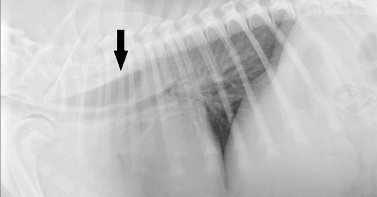
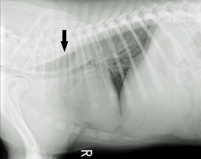
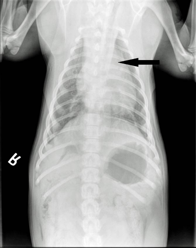
Radiographs of the thorax were performed (right lateral and dorsoventral; Figures 1 and 2, respectively).
A mild to moderate gas dilation of the cranial oesophagus was apparent at the level of the heart base/fourth intercostal space, just cranial to the heart base (Figure 1, black arrow). The caudal aspect of the oesophagus was normal.
From the dorsoventral view (Figure 2, black arrow), leftward tracheal deviation at the level of the cranial border of the heart was appreciated. No other mediastinal abnormalities were seen.
The lung fields showed normal opacity and volume, and no signs of aspiration pneumonia were present. The radiographic diagnosis was focal dilation of the oesophagus cranial to the heart base and left-sided deviation of the trachea.
These radiographic findings, together with the clinical history and patient’s age, were suggestive of vascular ring anomaly, causing constriction of the oesophagus and subsequent cranial dilation.
Given the leftward deviation of the trachea, persistent right aortic arch (PRAA) was very likely. Other possible, but much less likely, differential diagnoses were oesophageal stricture or an oesophageal foreign body.
An extraluminal mass, oesophageal neoplasia or gastric disease were not considered, given the patient’s young age.
Neuromuscular, endocrine or immune‑mediated disease were considered not likely due to absence of other accompanying clinical signs and because the oesophageal dilation was focal rather than generalised. Since a vascular ring anomaly was the main clinical suspicion, CT of the thorax with angiography was performed and confirmed a PRAA with no other concurrent vascular anomalies.
Acquired oesophageal obstructions due to vascular ring anomalies are a common cause of regurgitation in young dogs. Different types of vascular malformations from the embryonic aortic arch may occur alone or in combination, and may or may not be of clinical significance.
PRAA, aberrant right or left subclavian artery, double aortic arch, right patent ductus arteriosus, persistent right dorsal aorta and aberrant intercostal arteries are the main congenital vascular anomalies in dogs. PRAA accounts for 95% of all vascular ring abnormalities seen in dogs.
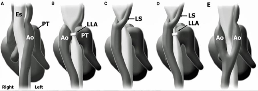
Surgical treatment is recommended and the overall long-term outcome is good or excellent for 87% of dogs. An 8% mortality risk exists prior to discharge and 18% after. Due to the possible combination of different vascular abnormalities (Figure 3), CT angiography is recommended preoperatively.