12 Sept 2022
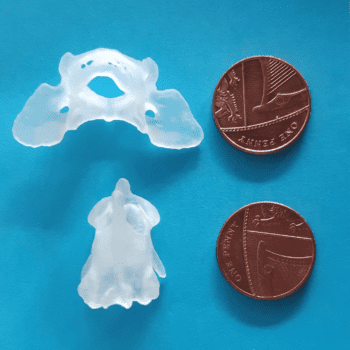
Lorenzo Mari and his team modified a dorsal AA stabilisation technique by combining it with the use of 3D printed guides to maximise precision of screw placement in Yorkshire terrier.
James Westgate
Job Title
Dotty the Yorkshire terrier, seen here awaiting surgery, was walking just two days later.
The Ralph’s neurology team has added a unique modification to a novel dorsal atlantoaxial (AA) stabilisation technique to maximise the precision of screw placement.
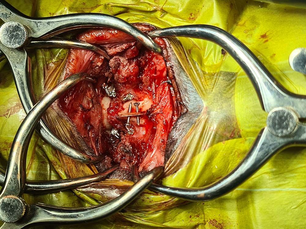
The technique has now been successfully performed on two canine patients, including a Yorkshire terrier called Dotty, who was referred to The Ralph’s neurology and neurosurgery team following sudden paralysis in all four of her legs.
A full neurological examination, including an MRI and CT scan, on the 2.4kg dog confirmed the suspected diagnosis of AA subluxation.
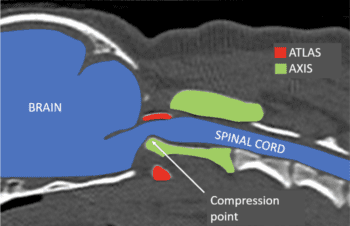
AA subluxation is a condition most common in “toy breeds” where the first two vertebrae of the neck (the atlas and the axis) subluxate (misalign), which traumatises and compresses the spinal cord.
The condition can be congenital or traumatic, and can present in many ways – from episodic pain to sudden paralysis, respiratory difficulties and even sudden death.
The Ralph’s head of neurology and neurosurgery, Lorenzo Mari, operated on Dotty with a novel dorsal AA stabilisation technique first published last year by Guillaume Leblond et al.
Mr Mari and his team modified the technique by combining it with the use of 3D printed guides to maximise the precision of screw placement in each individual case.
Dotty was walking again just two days after surgery while a postoperative CT scan showed normal alignment of the atlas and axis, with the screws positioned perfectly and the spinal cord decompressed.
Mr Mari said: “I was lucky enough to work with Dr Leblond last year and learn this technique directly from him. Dorsal stabilisation techniques present very many advantages compared to the most classically used ventral techniques.
“Not only do they better respect the biomechanical principles of fracture/luxation stabilisation, but most importantly, they avoid dissections close to extremely important and delicate organs, such as the larynx, the pharynx, and important nerves and vessels, minimising the risk of serious complications related to postoperative dysfunction of these structures.”
VIDEO: Lorenzo Mari realigining Dotty’s axis to its correct position during dorsal atlantoaxial stabilisation surgery.
Mr Mari added: “The dorsal technique allows much more space to place the surgical cement that joins the screws together, which can also cause mechanical compression or irritation to the above listed structures when placed ventrally.
“The problem of dorsal techniques has always been that the constructs previously described appear to be pretty weak compared to the ventral techniques, and this is where Dr Leblond’s technique changed the game, having the merit to identify safe and pretty generous bone corridors to introduce the screws.
“The unique modification that we made here at The Ralph, in collaboration with Vet3D, was to combine this technique with the use of 3D-printed customised guides that can maximise the precision in placing screws in each single case.
“We must not forget that most of these dogs are somewhere around 2kg in weight and we are talking about inserting 1.5mm screws in 2.3mm to 2.5mm bone corridors, where the margin for error is close to zero.”