18 Mar 2020
Video-assisted thoracoscopy and removal of part of the lung reveals the cause of patient’s abscess – a tiny, inhaled grass seed.
David Woodmansey
Job Title
Ruby underwent a video-assisted thoracoscopy to allow a visual of the whole of her chest cavity. Image: London Vet Specialists
A vet team has got to the root of a dog’s long-term lung infection after detecting a single grass seed lodged deep in the patient’s lung.
Ruby, the four-year-old Havanese, had been unwell for three months, struggling with a persistent, chronic cough and frequent high temperatures.
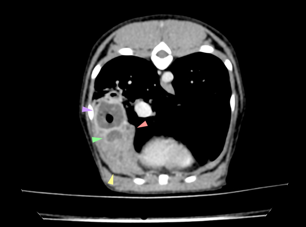
She was finally referred to London Vet Specialists (LVS) in Belsize Park, London, where, coughing and slightly feverish, she was sent for a CT scan and a bronchoscopy.
Internal medicine specialist Hannah Darcy said: “Images from Ruby’s scan showed an abscess in her right lung, while the bronchoscopy showed inflammation of her airways, with pus leaking from the entrance to one of the small airways.
“A sample of the infected fluid was taken to determine which antibiotics Ruby would require.
“However, the concern was that while antibiotics may prevent infection from spreading throughout Ruby’s lung, they would be unlikely to fully penetrate and treat the abscess. It was then decided to remove the infected portion of the lung to give her the best chance of a full recovery.”
Ruby subsequently underwent a video-assisted thoracoscopy (VATS) performed by LVS head of surgery Janet Kovak McClaran.
Dr McClaran added: “A camera was inserted into Ruby’s chest via a small incision between her ribs, which allowed me a full visual of the whole of her chest cavity. This allowed the infected lung lobe to be removed and the underlying cause of Ruby’s lung abscess was finally revealed – a grass seed.
“She must have inhaled it a number of months before, with it lodging in one of her small airways and causing an infection that would be impossible to treat without removing the offending foreign body.”
Ruby has made a rapid recovery from surgery, with her cough instantly cured.