12 Feb 2018
Francesco Cian, in this month’s Cytology Corner, looks at a case of cutaneous histiocytoma in a two-year-old cocker spaniel.
Francesco Cian
Job Title
Figure 1. An aspirate of the lesion.
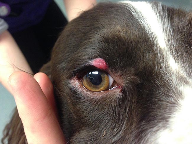
Figure 1 is a picture (Wright-Giemsa, 50×) from an aspirate of an upper eyelid lesion (Figure 2) in a two-year-old cocker spaniel.
The submitted smear is highly cellular with adequate preservation. The background is basophilic with occasional red blood cells, bare nuclei and lymphoglandular bodies. The latter (green arrow) are cytoplasmic fragments from lymphoid cells.
A mixed population of nucleated cells is present, mainly histiocytic and lymphoid in origin. Histiocytes (red arrow) have moderate amounts of lightly basophilic to clear cytoplasm, with poorly defined borders, while nuclei are round, occasionally indented and, paracentrally located, with granular chromatin and poorly visible nucleoli. Anisocytosis and anisokaryosis are mild. Lymphoid cells are mainly small lymphocytes (black arrow); however, a few intermediate and large size elements are also seen.
These findings are consistent with regressing cutaneous histiocytoma.
Cutaneous histiocytoma is a benign neoplasm originating from epidermal dendritic or Langerhans cells. It often appears as a solitary, dome or button-shaped dermal lesion, variable in size, often bright red in colour, rapidly growing and prone to ulceration. It is typically found on the head (including the pinna) or limbs of young dogs, and certain breeds (for example, boxers and dachshunds) seem to be predisposed. However, histiocytoma may occur in dogs of all breeds and ages.
Cytologically, cutaneous histiocytoma is characterised by a proliferation of histiocytic cells, which may show features of atypia (including binucleation, anisocytosis/anisokaryosis and mitotic figures), despite its benign behaviour. In mature lesions, it is not uncommon to observe an infiltrate of small lymphocytes representing the T-cell immune response, often leading to a spontaneous regression of the lesion within a few months.
Occasionally, cutaneous histiocytoma may resemble tumours of other origin – in particular, lymphoma. This may be a concern for lesions appearing in adult/old animals. Therefore, presumed histiocytomas that do not resolve or increase in size should be biopsied and submitted for histopathologic examination to rule out cutaneous lymphoma.
Histiocytoma is almost always a solitary lesion and the existence of multiple histiocytomas at the same or distant sites is considered uncommon. When this occurs, the umbrella term canine cutaneous Langerhans cell histiocytosis (LCH) is preferred. Feline counterparts of canine cutaneous histiocytoma and cutaneous LCH have not been reported.