
15 Sept 2026
Study links faecal IgA levels with atopic dermatitis
Faecal IgA was found to be significantly lower in dogs with canine atopic dermatitis compared with healthy controls.

Chris Simpson
Job Title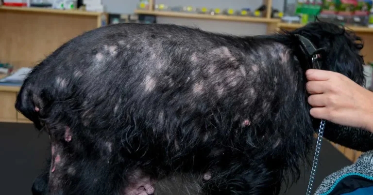
Image: Todorean Gabriel / Adobe Stock
Researchers have shed new light on a chronic skin disease in dogs after discovering it is associated with faecal concentration of a key antibody protein.
Scholars from the Royal (Dick) School of Veterinary Studies and Scotland’s Rural College (SRUC) found faecal immunoglobulin A (IgA) concentration was associated with canine atopic dermatitis (CAD).
The BSAVA PetSavers-funded study, published in the Journal of Small Animal Practice, characterised IgA levels in the faeces and saliva of dogs presenting to veterinary practice with CAD.
The researchers recruited 32 atopic dogs across 19 breeds through the Royal Dick’s Hospital for Small Animals dermatology service between November 2023 until March 2024.
They also recruited 36 largely breed-matched dogs through the hospital’s general practice as controls. Control dogs were at least four years of age with no history of skin disease and had not received immunosuppressive or other medications likely to affect immune function.
ELISA testing
ELISA (enzyme-linked immunosorbent assay) testing was performed to quantify mean IgA concentrations in the dogs’ faeces and saliva. Mean-ranked faecal IgA was found to be significantly lower in dogs with CAD than controls, while salivary IgA was not.
Faecal IgA concentrations in the atopic dogs ranged from 0.05g/L to 5.28g/L, while in control dogs it ranged from 0.014g/L to 10.27g/L, with a difference between medians of 0.803g/L. Saliva IgA concentrations in the atopic dogs ranged from 0.02g/L to 2.55g/L compared with 0.0012g/L to 2.82g/L in the controls, with a difference in the medians of just 0.108g/L. Faecal and salivary IgA were not correlated with one another in either the atopic or the control populations, nor were they correlated with age in either population.
No individual dogs in either group had both low faecal and salivary IgA, which the authors said suggested that none of the dogs had an IgA deficiency.
Build on evidence
It is hoped the paper will build on existing evidence that low IgA is associated with CAD, and the authors noted further work was required to determine the significance of low faecal IgA in the development of tolerance or hypersensitivity to food and environmental allergens.
They called for future research to be carried out on larger cohorts and on high-risk breeds from birth through to adulthood to increase understanding of the role of IgA in development of gut tolerance versus immune dysregulation.
The scholars added that further exploration of the interaction between host IgA, the gut microbiome, skin and the environment will help vets overcome challenges in determining the most effective treatment for individual dogs.
‘Beyond food allergy’
SRUC veterinary immunologist and corresponding author Francesca Soutter said: “This work reveals a potential role for gastrointestinal mucosal immunity in CAD, that may go beyond food allergy.
“We hope to explore and understand this association through further research to provide improved treatment and prevention strategies.”