22 Apr 2019
Ryk Botes looks at the development of this procedure by detailing the diagnosis, surgery and outcome in an English springer spaniel.
Ryk Botes
Job Title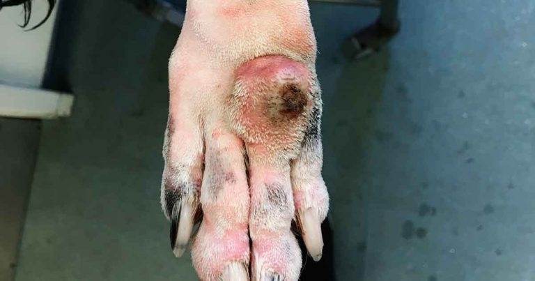
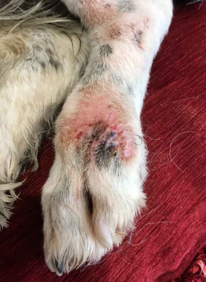
Figure 1. The granuloma.
Hygroscopic skin expansion has previously been used on the trunk, head and proximal limb of dogs, with considerable success.
However, until now, its application distal to the carpus and tarsus has been more complicated – presumably because the skin in this region is less robust than elsewhere and blood supply is easily compromised (De Lorenzi et al, 2018).
A recent adaptation of the protocol used in other anatomical areas has made it possible to expand tissue of the distal limb with minimal complications. This has been achieved through careful selection of tissue expander size, cautious placement in relation to the surrounding vasculature, and limiting expansion time to a maximum of 14 days.
The applicability of tissue expansion in this anatomical location offers a unique solution in cases that would, otherwise, require wound healing by second intention or free skin grafting.
To date, 11 dogs have undergone hygroscopic tissue expansion of the skin distal to the carpus and tarsus. The cases have been treated by surgeons of variable experience and most have been seen in first opinion practice – reflecting the fact implantation of the device and reconstruction are, in general, technically uncomplicated.
Of the 11 dogs that have undergone tissue expansion of the distal limb, primary closure of the deficit was achieved in 10 cases – giving a 91% rate of primary closure, suggesting tissue expansion is an effective method of creating tissue for the surgical repair of distal limb deficits in dogs.
One of the first dogs to undergo tissue expansion distal to the carpus was an 11-year-old male English springer spaniel with an acral lick granuloma over the fourth and fifth metacarpals.
A presumptive diagnosis of acral lick granuloma was made based on the case history and presenting signs. The dog had a three-year history of pruritus – thought to be the result of atopic dermatitis – and was intermittently managed with oclacitinib, lokivetmab and intermittent co-amoxiclav, marbofloxacin or clindamycin for secondary infection.
Persistent licking of the left dorsal metacarpal region was assumed to be secondary to atopy, but likely to have a behavioural component associated with attachment to the owner. Behavioural modification was attempted using the selective serotonin reuptake inhibitor fluoxetine, but to no avail. Laser therapy was used as an adjunct to stimulate blood flow to the area.
As the granuloma was refractory to medical management, surgical removal was elected. However, due to the size and location of the granuloma, it would not have been possible to achieve primary closure following excision without the use of skin expansion.
The dog was anaesthetised using a standard protocol and the surgical site prepared routinely. An incision was made on the lateral aspect of the limb, proximal to the granuloma. The SC tissue was undermined to create a pocket in which to sit the expanders.
Two hygroscopic skin expanders were placed in the created pocket – measuring 5mm × 27mm and 3mm × 22mm, respectively – and positioned against the margin of the granuloma (Figure 1).
Where expanders are used to create additional tissue ahead of oncological resection, they are placed outside the tumour margins to prevent seeding of neoplastic cells.
The wound was closed with a continuous layer of SC sutures, followed by a row of skin sutures.
The limb was left without a dressing to allow for continuous monitoring, while a buster collar was supplied to prevent interference. The dog was discharged the same day.
As with all dogs undergoing tissue expansion, analgesia was provided for postoperative pain for the first few days following implantation of the device, but continuation of this throughout the expansion process was unnecessary. In this case, the surgeon used meloxicam.
As with implantation of tissue expanders in other anatomical locations, the dog remained comfortable throughout expansion and did not show signs of lameness. Given the nature of the lesion, postoperative antibiosis – clindamycin – was provided to mitigate infection risk.
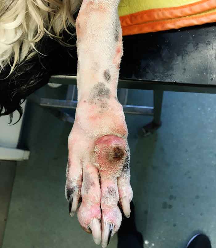
The dog was monitored by the owners at home, who were made aware of signs of infection, tissue necrosis and impediment of venous return. At 14 days post-implantation, expansion was complete (Figure 2).
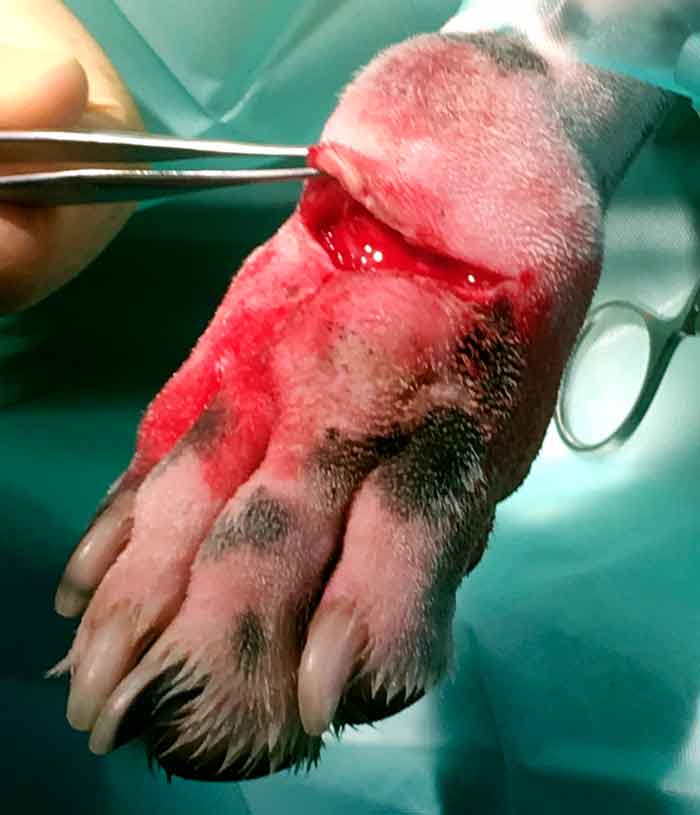
At 17 days post-implantation – 3 days later than recommended – the dog was re-anaesthetised for reconstruction. As a result, slight discolouration of the skin overlying the expanders was present, but this did not affect the viability of the skin – or its use in reconstruction of the wound – and resolved within 48 hours. The granuloma was resected and the expanders removed through the defect created (Figure 3).
The additional skin created during the expansion process was undermined and pulled distally to close the wound at the site of the excised granuloma, with minimal tension.
Despite having plenty of additional tissue, the shape and orientation of the granulomatous mass meant it was not possible to close the very lateral edge of the wound with the expanded skin. Therefore, a small amount of tissue was harvested from the lateral aspect by undermining of the SC fascia and pulling the liberated skin medially. Primary closure was achieved and the skin closed with interrupted sutures.
The dog was discharged with analgesia, meloxicam, and antibiotics – clindamycin and co-amoxiclav – and monitored postoperatively. No postoperative complications were observed.
At 14 days after removal of the mass and reconstructive surgery, the wound had fully healed (Figure 4). The sutures were removed and the dog was able to resume normal activity, with no reported reoccurrence of the problem to date.
The distal limb is a common location for injury, and frequent site of tumour and cyst occurrence. Limited, poorly elastic and thin tissue in this area makes wound closure extremely difficult, and wound closure under tension can lead to severe complications arising from circulatory compromise – resulting in tissue necrosis in the worst cases (Pavletic, 2005).
Limited tissue at this location makes the creation of tissue flaps virtually impossible, and free skin grafts from the thorax and abdomen are often fraught with complications (Lemarié et al, 1995). A major retrospective study of skin grafting suggests a graft survival rate of only 38% in dogs (Riggs et al, 2015).
Primary or delayed primary closure is preferable to healing by second intention – not only because of the additional time taken and increased infection risk associated with secondary wound healing, but because wounds that heal by contraction and epithelisation result in excess or fragile scar tissue that fails to protect the limb from subsequent injury and may lead to future complications (Tsioli and Dermisiadou, 2018).
Additional drawbacks include the need for repeat bandaging – often for several weeks – and the accompanying cost and management implications (Pavletic, 2005).
Hygroscopic skin expansion allows additional tissue to be created to achieve primary closure in locations where this would have, otherwise, been impossible or where wounds were at risk of breaking down due to excessive tension.
This technique lends itself particularly well to the reconstruction of wounds on the distalmost aspects of the limb, because of a lack of robust tissue in this location, and because this area of the limb is commonly affected by injury and pathology requiring surgical treatment.
The efficacy of this procedure is demonstrated by the successful reconstruction of distal limb wounds in 10 dogs following skin expansion. Despite the requirement for patients to undergo general anaesthesia, the process offers a practical solution because these animals would, otherwise, require lengthy management of open wounds.
The stress of repeat bandaging and discomfort of second intention wound healing can be avoided by opting for skin expansion and primary wound closure. An additional advantage is the reduction in scar tissue compared with secondary wound closure, which may reduce the chances of future injury at the surgical site.