11 Sept 2017
Kate Parkinson reports on a case study of a feline with a grass seed in its urethra, from its discovery to treatment and removal.
Kate Parkinson
Job Title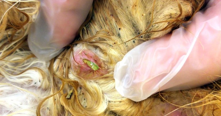
Figure 1. A grass seed protruding from Freddie’s urethra.
A cat presented with dysuria and was admitted for presumed urinary obstruction. Following anaesthesia, a grass seed was discovered partially blocking the cat’s urethra. Following removal of the grass seed and treatment of an ascending urinary tract infection, the cat made a full recovery. Urethral blockage caused by a foreign body rather than a urolith is an unusual, but important differential diagnosis for cats presenting with urethral obstruction. The grass seed in this case was tentatively identified as a barley grass awn (Hordeum species).
Grass seeds are common foreign bodies worldwide and may lodge in a variety of locations, including the urinary bladder. Grass seed foreign bodies are more common in dogs than in cats and the most common location for grass seeds is the external ear canal. In this case, the grass seed was easily removed; however, grass awn localisation may often be challenging. Seeds may be located via exploratory surgery, ultrasonography and CT.
A three-year-old male neutered cat, Freddie, presented with dysuria and pain on abdominal palpation.
The previous day, the cat had been treated by an out-of-hours emergency provider for sudden onset dysuria and a suspected partial urethral blockage. The notes from the emergency provider indicated the cat began to drip urine following admission.
A minimum database and urinalysis had been performed at the emergency hospital. A urine dip stick was positive for blood, protein, and white blood cells, and blood sampling revealed a slightly raised PCV and urea. As the cat was eating and seemed to be passing urine, conservative treatment of diazepam, buprenorphine and IV fluids were elected, and he was transferred back to the owner in the morning. Freddie presented to the general practice that same morning.
Freddie is a domestic longhaired cat with no history of dysuria. On presentation, he was alert and responsive, but quieter than normal. His bladder was the size of a tangerine, and painful and firm on palpation. Urine dripped from his penis on abdominal palpation, and his rear end was matted and stained with urine. The patient’s prepuce appeared inflamed, although a detailed examination was not possible as he had become quite grumpy by this time.
A provisional diagnosis of suspected urethral obstruction with urinary overflow was made and Freddie was admitted for further investigation. Repeat blood sampling revealed a slightly low potassium of 3.4mmol/L (normal range 3.5mmol/L to 4.8mmol/L), a high normal urea of 12.3mmol/L (normal range 5.7mmol/L to 12.9mmol/L) and a slightly elevated creatinine of 237mmol/L (normal range 71mmol/L to 212mmol/L).
Although acute kidney failure and hyperkalaemia are common sequelae to urinary obstruction in cats, these abnormalities were absent in this case – perhaps due to the continued passage of small amounts of urine or previous IV fluid therapy. Freddie was anaesthetised intramuscularly with a premedication of acepromazine (0.01mg/kg) and buprenorphine (0.01mg/kg). Anaesthesia was induced with IV propofol given to effect. Following intubation, anaesthesia was maintained by inhaled isoflurane.
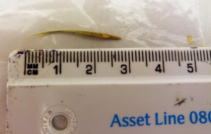
On exteriorisation of the penis, a piece of grass was found protruding from the urethra (Figure 1). Following gentle traction, a grass seed was removed, which measured 3cm in length (Figure 2), and approximately two-thirds of this had penetrated the urethra. After removal of the seed, the bladder could be manually expressed with moderate resistance. A sample of urine was collected via cystocentesis, and a tomcat urinary catheter passed easily without resistance. The patient’s bladder was emptied and flushed with 20ml of sterile saline.
After flushing the bladder, the urinary catheter was sutured in place and attached to a closed collection system. Unfortunately, the plastic collar of the catheter then detached from the catheter itself, necessitating removal and replacement of the catheter.
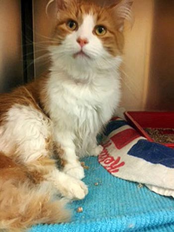
The anaesthetic was stable and Freddie recovered from his anaesthetic without incident (Figure 3). Following removal of the grass seed, meloxicam injectable suspension for cats 0.5ml (2mg/ml) was administered subcutaneously, together with 0.25ml amoxicillin/clavulanate (175mg/ml) to provide broad-spectrum first-line antibiotic cover pending culture and sensitivity results. IV fluid therapy was provided at a rate of 10ml/kg/hour.
The following day, Freddie was very bright and had eaten well. He had produced urine well overnight. The decision was made to remove his urinary catheter and he immediately urinated following removal. He continued to urinate well throughout the afternoon and was discharged the same day with a seven-day course of amoxicillin/clavulanate (12.5mg/kg) administered twice daily per os and a seven-day course of oral meloxicam administered at a rate of one unit per kg once daily per os.
In-house testing of the urine obtained via cystocentesis revealed a normal urine specific gravity of 1.035 and a trace of protein present on the urine dipstick. A sample of urine was submitted to an external laboratory. Urine culture revealed a >105cfu/ml bacterial count of Escherichia coli and Enterococcus species. Significant bacterial counts in cats with urine samples collected via cystocentesis were thought to be >103. Fortunately, both the E coli and Enterococcus species cultured were sensitive to amoxicillin/clavulanate, with a minimal inhibitory concentration (MIC) of <2.
The MIC of a bacterial culture is the lowest concentration of antibiotic that inhibits bacterial growth1. In practice, if the MIC for the bacterial isolate falls in the susceptible category, successful treatment (cure) is more likely than if the isolate were classified as resistant.
In this case, the results implied the antibiotic amoxicillin/clavulanate was effective at the lowest concentration tested and should, therefore, be an effective choice. The antibiotic in this case was not changed and Freddie was continued on twice daily amoxicillin/clavulanate for one week following his procedure.
Twenty-four hours following discharge, Freddie was still urinating well. His bladder was small and soft. He had urinated once on the kitchen floor, but was otherwise urinating normally with no sign of pain. His temperature was normal.
At a follow-up telephone call three days after discharge he was reported to have improved, although he had urinated once on the owner’s dressing gown when she left it outside on the lawn. He had also developed diarrhoea, thought to be secondary to the antibiotic use. Meloxicam was stopped, but the diarrhoea continued.
During a follow-up one week later, Freddie was much happier. His bladder was still small and soft, and he was eating and drinking well, although he had experienced three more episodes of inappropriate urination. A second urine sample was collected by ultrasound-guided cystocentesis. The ultrasound examination was unremarkable and urine culture was negative. Antibiotic therapy was discontinued. Freddie was switched to a canned diet to encourage urination and restarted on oral meloxicam. Two weeks later, Freddie continued to do well with no further episodes of inappropriate urination.
Differentials for feline urethral obstruction include uroliths that form in the bladder, but lodge in the urethra, and urethral plugs usually formed from an accumulation of cells, crystals and debris, or urethral spasm, which may be caused by inflammation or irritation of the urethra. Less common causes include urethral foreign body – as in this case – urethral stricture (usually secondary to trauma or inflammation) or neoplasia2.
Grass awns are a common seasonal problem in small animal practice worldwide and may be discovered in a wide variety of locations. Treatment of grass seed foreign bodies relies on successful and complete removal of the seed, which, in many presentations, may be challenging.
Grass seeds often cause foreign body reactions and have been recorded in the ears, paws, thoracic cavity3, bile duct4, eye, nose, lung5, prepuce, parotid duct6, vagina7, spinal cord8, and mediastinum9 of cats and dogs. In addition, grass seeds have been documented as a cause of meningoencephalitis10, endocarditis11 and iliopsoas myositis12.
Grass awns have been reported as causes of bladder foreign bodies in dogs13, where they are thought to migrate in a retrograde fashion from the urethral opening. Here the grass seed usually appears as a bladder stone or a linear hypoechogenic structure visible on bladder ultrasound14. The author found two anecdotal cases of grass seeds being lodged in the urethra of male cats reported on the message boards of the Veterinary Information Network. One case had the seed removed without incident. Unfortunately, the second case reported the grass seed was unable to be removed and the patient was euthanised due to financial concerns.
Grass seed foreign bodies seem much more common in dogs than cats. A retrospective review of 182 cases of grass awn migration in dogs and cats over a year found grass seed comprised 61% of all foreign bodies admitted15. Of the 182 animals admitted with grass seed foreign bodies, 8 were cats – 7 of which displayed ocular signs. Otitis externa accounted for 51% of cases. This reinforces the findings of a similar study carried out in the Riverina district of New South Wales that found otitis externa accounted for 47% of all grass seed foreign bodies seen in general practice16.
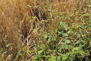
Grass seed identification was attempted using the online guide “Key to groups of British grasses”17, and tentatively identified as a barley grass due to the very long (>3cm) straight awns, possibly mouse barley (Hordeum murinum). This species is commonly found throughout England and Wales (Figure 4), and has been positively identified as a cause of cystolithiasis in male dogs and mediastinal abscess in a cat. Freddie’s owner reported he spent a lot of time outside and liked to spend much of this playing and sleeping in clumps of grass in the back garden, so it is not surprising he presented with a foreign body.
Although, in this case, grass seed removal was easy, diagnosis and removal of grass awn foreign bodies may often be challenging. Grass seeds in easily accessible locations, such as ears and eyes, may be relatively easily removed. Surgical exploration of an obvious discharging tract or swelling may be carried out, but may, in some cases, be unfortunately unrewarding. Grass seeds are rarely located on radiography, but may be located via ultrasonography (for example, in the bladder)18, radiography19 or CT scanning20.
This was an interesting case with a good outcome that required little long-term management following grass seed removal. Although the author has removed numerous grass seeds from the ear, eye, skin, interdigital web and feet of dogs, this was the first time experiencing a grass seed in the penis of a cat.
Grass awn migration up the urethra into the bladder, although theoretically possible, fortunately, did not occur in this case. If the grass seed had become lodged further up the urethra, it could have caused a complete blockage, which would be relatively difficult to remove and, unlike urinary calculi, would have been undetectable on radiography. Fortunately, in this case, the grass seed was relatively easily retrieved and Freddie made a full recovery following treatment of a secondary urinary infection.
The author would like to thank the staff of Orwell Veterinary Hospital, Freddie’s owner for permitting publication of this case – and photographs – and Katherine Ashton BVSc, MRCVS for reviewing the article.