13 Mar 2017
Victoria Brown recalls the search for a missing uterine horn during an irregular kitten spay.
Victoria Brown
Job Title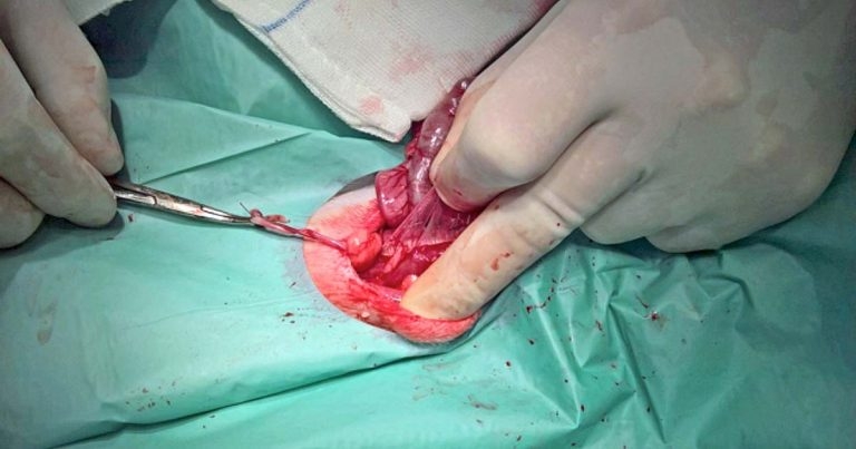
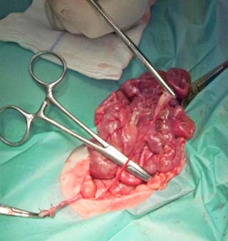
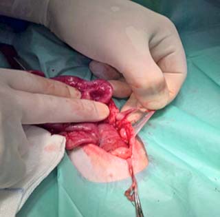
Figure 1. The single uterine horn and its ovary have been retracted.
A very cute four-month-old kitten presented for spaying.
I opened her up in the standard left flank approach, fished around with a spay hook, found a uterine horn and ovary, pulled them out (Figure 1), ligated, and cut them away from the body wall.
I traced the uterine horn back to the uterine body, but where was the second uterine horn? Vanished, invisible, missing, presumed dead?
Obviously, I was doing something wrong. I pulled on the uterine body, but no go. I pulled harder – any harder and the whole thing would tear off.
Thankfully, the clinical director was around (on his day off) and offered to dive in, remarking: “I’ve seen two of these before.”
Really? Perhaps he spends far too much time neutering.
He went “fishing”. “Uterus unicornis,” he pronounced. “This kitty has only one uterine horn. But they almost always have two ovaries. So we need to find it and whip it out, otherwise she’ll still come calling, even if she can’t get pregnant.”
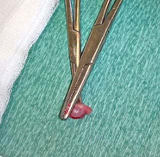
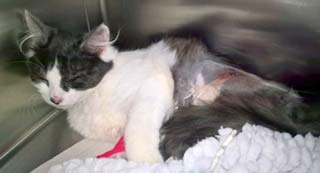
The kitten’s left flank incision was sutured, she was rolled into spinal recumbency and a midline approach made. Quite clearly, the uterus led to a cervix without forking off first into another horn. Where the horn should have been was a membranous ligament, delicate as a cobweb and leading to a full-sized ovary (Figure 2). The ovary was ligated and removed (Figures 3 and 4), the kitten was put back together (Figure 5). She was later bouncing about, oblivious to her unusual anatomical history.
Reading up on this wonderfully named condition later, with visions of unicorns and uteri floating through my head, I found no known scientific study exists to prove uterine unicornis is a hereditary genetic disorder.
The condition can be detected by x-ray or ultrasound prior to spaying if the patient has a family history of the medical condition (and if you’re good at reading radiographs/scans – I’m not). In some cases (approximately 50%), cats may also be missing a kidney on the same side as it’s missing a uterine horn (unilateral renal agenesis).
Apologies to those who know about this condition, but if there are any who don’t, I thought it was worth a share.