22 May 2024
Team at Aura Veterinary and a consultant from Royal Surrey County Hospital perform treatment typically used for people on 14-year-old dog.
Paul Imrie
Job Title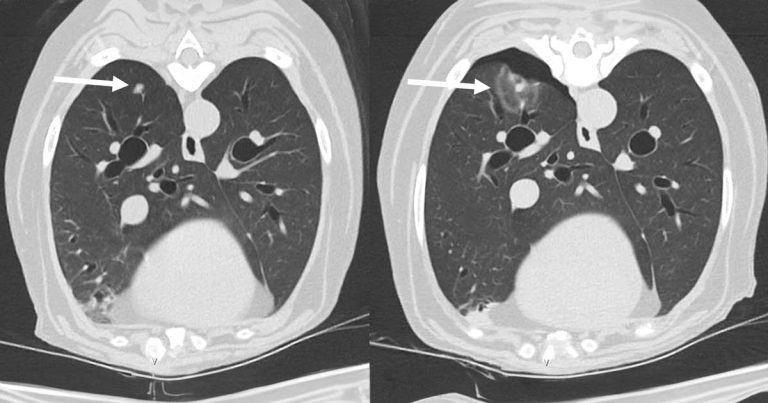
The 7mm lung nodule on a CT scan before (left) and immediately after (right) microwave ablation showing a sphere of ablated tissue and small pneumothorax.
Clinicians at Aura Veterinary teamed up with a consultant from a human hospital to carry out a cancer treatment routinely used in humans on a dog.
The 14-year-old female neutered Belgian shepherd had microwave ablation (MWA) – a minimally invasive treatment – on a lung nodule.
The dog had limb-sparing surgery three years previously for a large SC histiocytic sarcoma over the carpus, followed by a course of lomustine chemotherapy.
It developed slowly progressive metastatic disease 18 months later, which was managed with minor surgery to remove lymph nodes and chemotherapy, but a solitary slowly growing 7mm nodule in the right caudal lung appeared resistant to the treatment.
Aura Veterinary oncologist Quentin Fournier and radiologist Sergio Guilherme worked with Alex Horton, human interventional radiologist at the Royal Surrey County Hospital, to treat this lung lesion with MWA.
For the treatment, the tip of a special needle a few millimetres wide was inserted into the lung nodule under CT guidance, which was headed to create a sphere of thermal ablation that destroys the target mass.
The dog recovered well, with no further complications, although it did die six months later due to kidney disease.
Aura Veterinary said MWA can be used in veterinary medicine as part of a multimodal treatment plan, and ideal candidates should have a limited number of small nodules away from large vessels or other vital structures.
MWA is used in human medicine to treat breast, liver, lung and kidney tumours, and previously in dogs outside the UK in the liver, lungs, bones and kidneys.
Vets who may have a potential case can email Aura Veterinary.