18 Jun 2018
Ross Allan, in the first of a two-part article, describes wound healing as a journey – along which the owner should be fully engaged.
Ross Allan
Job Title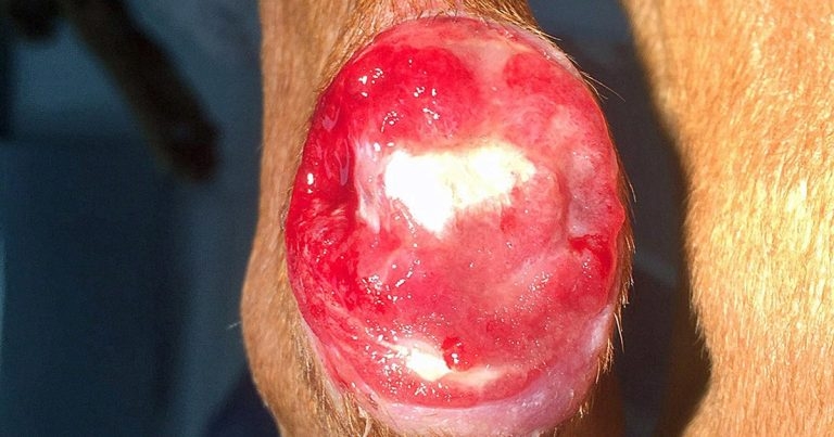
Wounds are something we encounter every day in small animal practice, and while most will heal with minimal complications, others can be immensely challenging to manage. Careful consideration must be given to each of the elements that contribute to successful wound healing and appropriate strategies must be put in place to manage these cases effectively.
This article recaps the classification of wounds and their pathophysiology and clinical management, as well as exploring the elements of client management, which can be important when dealing with such injuries. While accurately predicting the “journey” any wound takes is difficult, proper communication, an understanding of clinical concepts, case continuity, and careful case management can turn potentially frustrating and upsetting cases into some of the most satisfying you may deal with in practice.
Wounds occur in all manner of shapes and sizes – something we encounter and create almost every day. Most heal without incident or concern, but wounds are also presented that give us sleepless nights – wounds that won’t heal, are in tricky places or in difficult-to-manage patients.
We should all strive to improve our management of such cases from start to finish, and this article seeks to outline steps that can be taken to achieve “closure”.
Four key physiologic steps to wound healing exist:
1. Clotting phase – the formation of a fibrin clot at the site of injury.
2. Inflammatory phase – recruitment of white blood cells to protect the site of injury.
3. Proliferative phase – neovascularisation and cellular proliferation.
4. Maturation phase – remodelling and adaptation of tissue.
These stages occur in a predictable order and time frame after the initial injury, with the immediate initial clot forming due to platelet contact with collagen inside the vascular tissue and leukocytes being chemotactically drawn to the area (first polymorphonuclear cells, then mononuclear cells) over the initial three to five days.
The endothelial and fibroblast proliferation correlates with the development of granulation tissue at the wound site from day three to five onwards and, when a sufficient granulation tissue bed has developed, epithelialisation and wound contraction can start to occur. Eventually, as time progresses, collagen tissue is deposited by fibroblasts and this remodels to strengthen the wound. This phase can take weeks to months.
These four stages are similar to the other commonly used description of wound healing, which identifies the phases as inflammation, debridement, repair and maturation.
When faced with a tricky wound it is important to think about these discrete stages and at what point in the “journey” the wound is at. This will allow the clinician to identify the most appropriate measures that can be used to further assist and promote effective healing.
It is useful to consider the differences in wound healing described in dogs and cats, with some studies showing granulation occurs slower in cats compared to dogs. Also, while in dogs granulation tissue forms across the entire wound bed, in cats it progresses more circumferentially. A difference also exists in wound contracture, with cats relying more on myofibroblasts for wound contraction, whereas dogs rely mainly on migration of epithelial cells across a healthy granulation bed. These factors can be important to consider when managing wounds – similar wounds can behave differently, based on location, orientation and species.
Before exploring what can be done to support wound healing, it is important to review the classifications. Wounds can be split into the following main groups (Balsa and Culp, 2015), depending on the closure type that can be used:
While the four described classifications cover most types of wounds, some do not involve the entire thickness of the skin – resulting in partial epidermal/dermal destruction. Examples of these include skin burns – either thermal or chemical. In these situations re-epithelialisation can develop from the surviving epithelial tissue, meaning hair growth can occur and scarring will be less common than in wounds that heal by secondary intention. Wounds that heal through adnexal re-epithelialisation will not generally require intensive surgical management, but debridement may often still be necessary.
All of us in practice will know large wounds are a journey, and wound management is fraught with its ups and downs – dressings falling off, bad smells, chaffing and other challenges.
Educating owners on what to monitor for when their pet is having complex wounds managed is essential from the start. Ensuring owners are aware problems are likely to be anticipated during healing and providing them with an appreciation of the stages of healing can, in the author’s opinion, go a long way to keeping them onside throughout what is often a long and potentially arduous process.
Some tips to ensure owners remain engaged in the healing process are listed in Panel 1.
Additionally, one of the challenges is the healing process is dynamic and the treatment plan changes. While secondary closure may often be an option, a number of factors may influence the decision on whether to perform this – notably, classification of the wound, contamination, status of vascular supply and likelihood of unintended consequences with continued open wound management (for example, contracture over joints). These factors, as well as financial pressures, mean it is wise for clinicians to counsel owners of the advantages and potential risks of secondary closure should it be considered during the wound healing process.
Primary wound closure has been described as being unachievable in six main wound types:
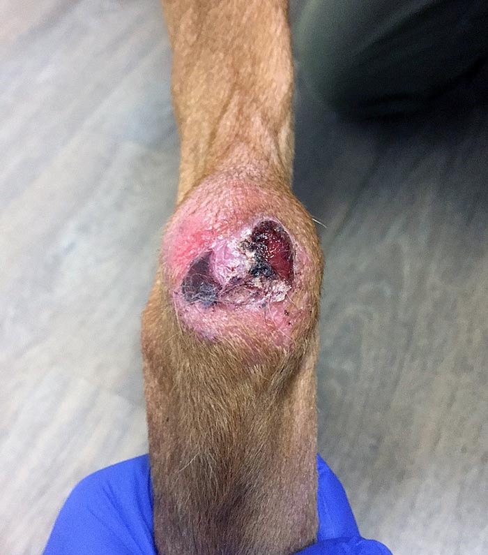
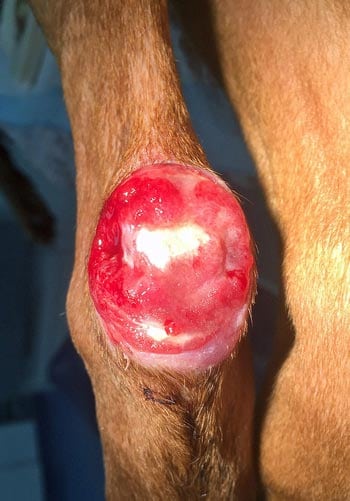
Case study 1. A decubital ulcer in a two-year-old pharaoh hound
Decubital ulcers (note these are more accurately named pressure ulcers, as decubital suggests the patient lies down, but they can also occur if able to stand) occur due to pressure over bony prominences causing ischaemia of the skin, full dermal loss and, often, bone exposure. They can also be caused by external splints and dressings that have been applied too tightly, and are more common in debilitated or paralysed patients, those of heavier weight and breeds with thin skin.
The sites most at risk of developing pressure ulcers are the greater trochanter, calcaneal tuber and olecranon. Grade one to four ulcers can develop. Managing these ulcers can be especially challenging and the author suggests reading the description by Pavletic (2018), which provides further detail on their classification and management strategies.
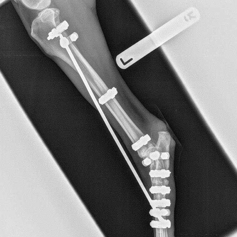
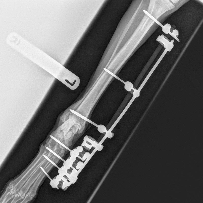
These can be very challenging wounds to manage, and steps to minimise direct pressure – perhaps through the use of “doughnuts”, and secondary dressing-related dynamic wound abrasion through the use of transarticular external fixators – can be useful tools to allow these to heal. In this patient, the decubital ulcer healed via secondary intention healing.