9 Sept 2025
Kayleigh Cook CertAVP(ESST), PGCertVPS, PGCAP, FHEA, MRCVS discusses what to look for and questions to ask owners to glean information required to make the best decisions in these pressured cases
Kayleigh Cook
Job Title
Image: Alexia Khruscheva / Adobe Stock
Attending a horse with colic in a field situation is one of the most frequent, and often challenging, emergency consultations in equine practice.
Owners are often very stressed, due to the perceived nature of the poor outcome of disease, and horses themselves can often be severely distressed and, therefore, difficult to deal with.
Clinicians are often faced with multiple pressures, such as safety, finances and the need to decide on management quickly. However, if managed appropriately, they can often be some of the most rewarding cases to deal with in equine practice.
Many studies have been performed looking at risk factors for colic in populations of horses. Older horses and geldings have been found to be at increased likelihood of strangulating lipomas (Garcia-Seco et al, 2005), in comparison to younger horses which are at increased risk of idiopathic focal eosinophilic enteritis (IFEE; Archer et al, 2014).
Broodmares in the postpartum period and taller horses have been shown to be at increased risk of large colon volvulus (Suthers et al, 2013), whereas miniature ponies have been shown to be at increased risk of faecoliths (Miklavcic, et al, 2024). The location of the horse can sometimes also provide a clue to the type of colic present, with some conditions such as IFEE having increased risk in certain locations (Archer et al, 2014).
Both history and signalment can provide clues as to the type of colic you may be expecting to see in the horse you are attending.
By asking the owner a few simple questions on the phone, it gives you something to consider as you drive to the horse, allowing potential diagnostic steps to be considered before your arrival. Some key questions to consider are:
Duration and severity of signs can guide you towards a type of colic. Colic types with significant compromise in blood supply often present as acutely painful, whereas horses with tympanic colic may initially present with mild signs of pain, but pain levels may increase if gas continues to build up (Singer and Smith, 2010; Southwood, 2023).
Changes in management such as diet, stabling, turnout and exercise have been shown in several studies to be a consistent risk factor for colic across all types of horses (Curtis et al, 2019).
Pelvic flexure impactions are a significant risk in any horse moved suddenly from pasture to a stable and provided with dry roughage, such as when following an injury or the end of summer turnout (Hudson et al, 2001). Stereotypical behaviour such as wind sucking has been shown to be associated with increased risk of epiploic foramen entrapment (Archer et al, 2008). The presence of parasites or dental disease has also been demonstrated to be a consistent risk factor for multiple types of colic (Curtis et al, 2019).
Administration of medications such as NSAIDs increases the risk of disease such as right dorsal colitis which can lead to colic signs (Flood et al, 2023).
Colic is a non-specific sign and may be confused by owners. A good history can help you rule out other causes of disease such as non-abdominal pain and toxicities. In some cases, with an acutely painful horse, emergency treatment may be required, but a follow up detailed history can be of value to obtain once the horse is stable.
When performing your clinical examination, bear in mind the clues gained from your history, but try to avoid this clouding your judgment when performing the exam.
If it is safe to do so, a thorough clinical examination should be performed consistently on all colic cases. Some cases may be too painful to fully examine without some form of analgesia; this should be provided before continuing your examination, for both human safety and animal welfare reasons.
In some instances, initial clinical examination can be unrewarding; however, this assessment can form a baseline for repeated clinical examinations which can guide your understanding of changes in pain levels and clinical parameters over time. Key aspects of your clinical examination should include both a distant and hands-on examination.
A distant examination should include an appraisal of:
A subjective initial assessment of the level of pain is the key indicator to take from this aspect of the examination. A significant degree of pain remains the single most reliable indicator of surgical colic lesion and should not be ignored (Singer and Smith, 2010). A hands-on examination can then include some or all the following:
The cardiovascular examination will provide information on the hydration status of the animal and indicate if any signs of hypovolaemia or systemic inflammatory response syndrome (SIRS) are present. Strangulating conditions or lesions which cause a complete obstruction of the gastrointestinal tract rapidly lead to fluid sequestration into the intestine and signs of hypovolaemia. If the lesion also leads to ischaemia, then signs of SIRS such as congestion or discolouration of the mucus membranes will occur.
Heart rate is a key indicator of pain, but will also increase with hypovolaemia in an effort to provide adequate tissue perfusion (Singer and Smith, 2010). Therefore, an animal with tachycardia (more than 60bpm), alongside other indicators of hypovolaemia such as dry or tacky mucus membranes, should increase suspicions of a more serious disease process. Respiratory rate will increase with pain and stress, as well as with increased abdominal distension in tympanic conditions.
Auscultation of abdominal borborygmi will give an indication of intestinal motility – strangulated portions of intestine will have no audible motility, whereas hypermotility should be expected in cases of spasmodic colic. Rectal temperature can be useful to differentiate horses with an inflammatory or infectious disease from those with colic (Southwood, 2006).
It also serves as a useful test of the horse’s demeanour prior to a rectal examination.
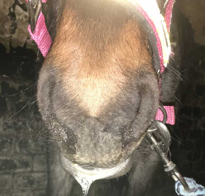
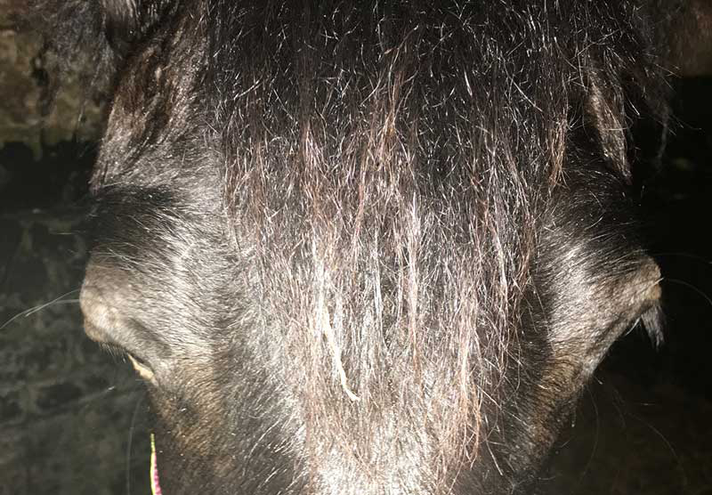
It is also important to consider other factors that may suggest other causes of disease which may present with similar signs to a colic episode – an example of this is equine grass sickness which, in the acute form of the disease, will often present with colic signs, as well as additional signs such as salivation, muscle fasciculations and ptosis (Figure 1 and 2; Wylie and Proudman, 2009).
Rectal examination is a useful diagnostic test to evaluate horses with colic, despite only 25% to 30% of the abdomen being palpable. It is often one of the most helpful tests for determining if a surgical lesion is present.
Human and animal safety are a vital consideration before attempting the procedure, and adequate restraint of the horse must be provided. A careful systematic palpation should allow clinicians to evaluate the abdomen for the presence of distended large or small intestine, impacted large intestine, abnormally located viscera or abnormally palpable taenial bands and masses. Multiple loops of distended small intestine or severely distended large colon may prevent a full rectal examination.
Detection of displaced abdominal viscera, abnormal orientation of taenial bands, oedema of the intestinal wall or distension of viscera, in particular small intestine, are abnormalities that may indicate a surgical lesion. Spasmodic colic often presents with no specific abnormalities palpable on rectal exam (Singer and Smith, 2010). Rectal examination findings should be considered alongside the rest of the examination.
The key question you need to answer by the end of your examination is: does this case require medical or surgical treatment?
While a specific diagnosis may not have been reached, this is not a primary concern if a horse is clearly a candidate for surgical treatment, as any delay to providing this treatment is likely to markedly worsen the prognosis for the horse; therefore, if surgery is an option for the equid, then referral should be initiated without delay. In these cases, it is advisable to pass a nasogastric tube to allow for gastric decompression prior to referral, in case of significant gastric reflux.
The presence of gastric reflux is strongly suggestive of a small intestinal cause of colic. Very painful cases may also require sedation for additional analgesia during transport; however, gastric decompression can also improve comfort levels of horses with small intestinal lesions (Singer and Smith, 2010).
In cases where surgical treatment is not an option, depending on the severity of the signs, further treatment or diagnostics can be attempted; however, if indicated, euthanasia should be provided without delay.
Most cases seen in first opinion practice will present with mild clinical signs with an unremarkable clinical examination and will often fully resolve following analgesia.
However, some cases will present with mild abnormalities which are not severe enough to warrant immediate referral. In these cases, administration of analgesia can be useful to then allow further diagnostics to be performed. A poor response to analgesia can also be useful to indicate that more intensive management and treatment of a case may be required (Singer and Smith, 2010).
Basic blood analysis can provide additional information and guide treatment in a colic patient. Packed cell volume (PCV) and total protein (TP) give an indication of an animal’s hydration status, as well as guiding towards the likelihood of a surgical lesion. Animals with surgical lesions will often have increased PCV and decreased TP, indicating significant dehydration and protein loss due to SIRS (Singer and Smith, 2010; Southwood, 2006).
Lactate can be measured via a stable side machine, allowing a rapid assessment of tissue perfusion.
A blood lactate value of more than 2mmol/L should prompt consideration of a more serious form of colic (Southwood, 2006).
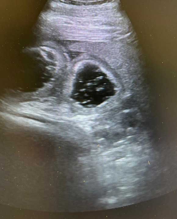
Abdominal ultrasound can be a highly useful non-invasive test to evaluate horses presenting with colic signs. Ideally, a 2.0MHz to 3.5MHz linear or sector ultrasound probe should be used for appropriate depth penetration (Singer and Smith, 2010). However, in emergency cases when the author has only had a 6.5MHz linear rectal probe in the car, confirmation of distended loops of small intestine has been achieved by utilising this probe to scan the inguinal regions. While this will not rule out distended loops of small intestine (Figure 3) if none are visualised, confirming their presence confirms the need to consider referral.
Ideally, the coat should be clipped to facilitate ultrasonography, but in an emergency case, the use of surgical spirit poured onto the coat can help to improve probe contact (Singer and Smith, 2010). Various techniques for abdominal ultrasonography are described in the literature, and the author uses the fast localised abdominal sonography of horses protocol to ensure a thorough and concise examination of the abdomen for common cases of colic (Busoni et al, 2011).
Abdominocentesis can be a useful procedure in cases with equivocal findings. It is a simple procedure to perform in the field, but does present risks such as intestinal laceration and peritonitis (Southwood, 2006).
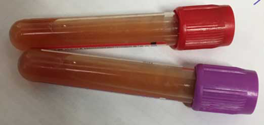
Normal peritoneal fluid should be clear and very light yellow in colour. Grossly, any sample that is cloudy, or discoloured pink to red, can suggest a more serious issue (Figure 4). Lactate, total cell count, and TP can also be measured. Increased peritoneal lactate concentration can provide an indication of ischaemia in the abdominal cavity – particularly if it is raised above the systemic lactate concentration (Southwood, 2006).
Following the additional steps performed, you should now have a greater confidence to decide if your case requires medical or surgical management. If surgery is indicated, then prompt referral should be arranged.
Some further medical treatment can be provided in the field if indicated; some examples of this include analgesia, antispasmodics, withdrawal of food or provision of very wet feeds.
Oral fluids via nasogastric tube can be very useful to treat large colon impactions. Light exercise such as lunging or in-hand walking can be beneficial in some cases of large colon displacements. In some medical colics, more intensive management may be required that is difficult or impractical to utilise in the field; therefore, hospitalisation of these case may be required.
The key question which needs to be answered promptly in all cases of equine colic is whether the horse has presented with a surgical lesion.
A thorough history and clinical examination are the key initial steps required to answer this diagnostic question. Specific indicators that would suggest a surgical lesion include severe signs of pain, tachycardia, tachypnoea, tacky or discoloured mucus membranes, increased PCV, absent abdominal borborygmi, significant abnormalities on rectal examination, poor response to analgesia or abnormalities discovered on ultrasonography, or on analysis of blood or peritoneal fluid.
Mild cases may require little more than adequate analgesia to ameliorate clinical signs. In many cases, an exact diagnosis will not be reached during the work up; however, a lot of very useful information can still be gathered.