
13 Aug 2026
Investing in diagnostic imaging equipment can transform patient care, but without clear clinical intent, it risks becoming an expensive liability. Practices must align hardware selection with actual caseload and team expertise, prioritising training and workflow integration to ensure measurable value…

Matt Winter
Job Title
For most first opinion practices, radiography and ultrasound are the mainstay of diagnostic imaging.
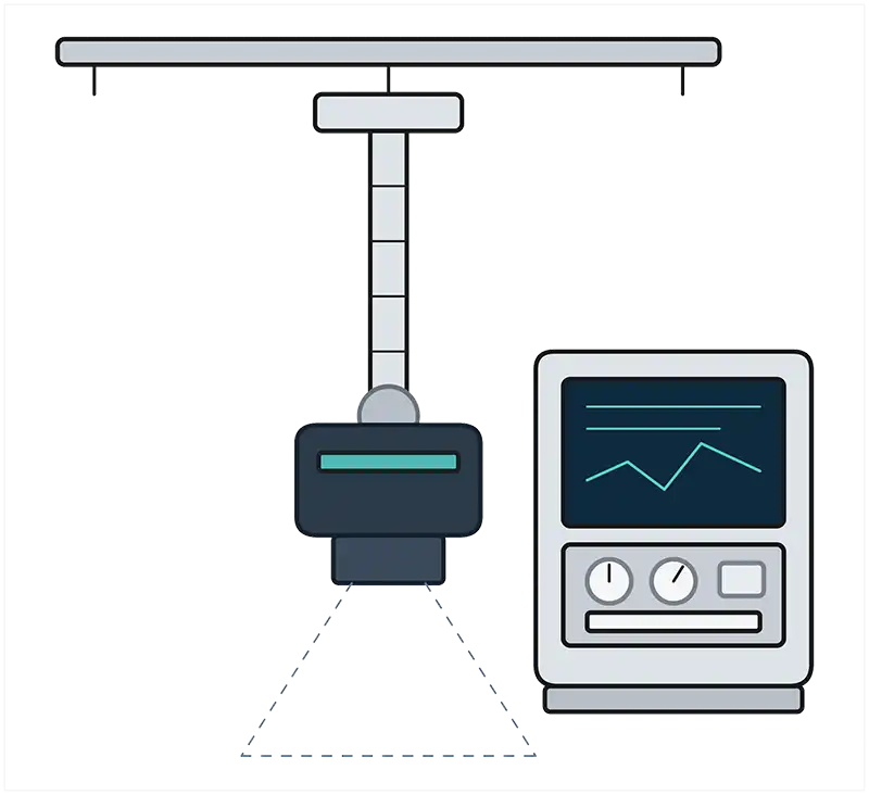
Digital radiography supports a broad general caseload, used for accurate screening of the thorax and abdomen, orthopaedics, trauma, emergency work and pre-referral decision-making.
Image quality depends not only on the generator, but on the detector, positioning aids and technique, exposure settings, and factors such as the practice culture and staff confidence. A high-quality digital plate is worth prioritising.
For orthopaedic cases, a mobile or rotatable tube head and a detector that is not fixed into one position can make appropriate projections and positional radiography easier, safer and faster.
Intra-oral dental x-ray is now central to good small animal dentistry, particularly in cats. A dental radiography unit should be considered essential for practices doing regular dentistry. It gives clinicians greater clarity, particularly in extraction planning and identifying retained roots, improving outcomes for patients.
Ultrasound is highly operator-dependent, both in acquisition and interpretation. Buying a better machine does not automatically create better ultrasound studies without the necessary human expertise. Probe choice is also a key investment consideration, and should be informed by caseload.
For small patients, a microconvex and linear probe may be sufficient. Larger dogs may require a lower-frequency curvilinear probe. Echocardiography typically needs a phased-array sector probe, continuous ECG display, appropriate measurement packages and specific training.
A practice should be realistic about how it will use the machine, not how it hopes it might use it. For many teams, it is better to build competence in abdominal ultrasound before buying additional equipment to master echocardiography.
With all modalities, PACS and PMS integration can markedly improve workflow, but poor integration can make everyone’s day harder.
Choose systems with strong DICOM compatibility, robust PACS and PMS integration, transparent data ownership, good export functionality, cybersecurity safeguards and supplier support for software upgrades.
Avoid being locked into proprietary formats that make second opinions, teleradiology, AI integration or future migration difficult.
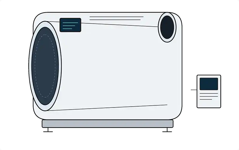
CT and MRI sit in a different investment category, yet the aforementioned principles remain. Purchase should be driven by actual caseload, not aspiration.
CT can be transformative for orthopaedics, complex trauma, surgical planning, oncology staging, nasal disease, neurologic disease, thoracic disease and selected exotic cases.
It is also increasingly useful in rabbits, where dental and skull disease can be difficult to fully characterise on radiographs. However, case selection is critical.
CT is not automatically “better” than ultrasound for abdominal patients, which is often less expensive, more targeted and often more useful, with less need for chemical restraint. CT also requires additional space, shielding and power requirements.
Practices considering CT should understand the distinction between cone-beam CT (CBCT) or flat panel CT (FPCT), and multidetector CT (MDCT).
CBCT/FPCT systems can be valuable for exotics species, including birds, reptiles and rabbit-sized patients, particularly for high-resolution skeletal or dental imaging.
They are, however, more susceptible to scatter and motion artefact and are not equivalent to the quality of MDCT for many soft tissue, contrast-enhanced or thoracoabdominal applications.
For general practice use, MDCT is almost always the more appropriate option.
MRI is even more caseload-specific. Its greatest value in small animal practice remains neurological disease, particularly brain and spinal cord imaging, as well as selected musculoskeletal indications.
Low and high-field systems are available. There are significant limitations to low-field systems. Specific space, shielding and power requirements must also be met. Clinically, the key question is not simply whether you can acquire MRI images, but whether you have the clinical expertise to act on the findings.
If a practice has no neurologist, no surgical pathway or no clear referral arrangement, MRI may generate diagnoses without solutions.
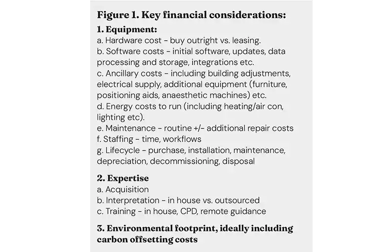
Hardware is only the start and the financial modelling must be rigorous. Practices need to consider purchase versus lease costs, installation, building work, shielding, electrical supply, cooling, anaesthetic equipment, contrast injector systems, software licences, PACS storage, maintenance contracts, insurance, downtime cover, staff training and eventual decommissioning or disposal. For CT and MRI in particular, service contracts can be a major annual cost (Figure 1).
With all systems – especially if refurbished – scrutinise the warranty carefully. Does the warranty include all the equipment? What is excluded? Is there a loan machine if yours fails? For ultrasound, transducers are expensive, fragile and critical to performance, so warranty detail matters. For CT, check whether the tube is covered; it is commonly one of the most expensive components.
Consider your realistic caseload – provided you have the right personnel and space within existing workflows. Do the financials stack up? If you need additional cases to justify the expenditure, ensure you have agreements with neighbouring practices or expansion plans to meet these estimates, while also ensuring that imaging is appropriate and clinically indicated.
Regulatory and safety requirements must be designed in from the start. Radiation safety should not be treated as a paperwork exercise. Veterinary use of radiation has expanded and protection should consider staff, members of the public, the environment and the animal patient. Imaging suite layout should allow staff to leave the controlled area quickly, use appropriate shielding and avoid manual restraint wherever possible.
CT typically delivers a much higher dose of radiation to the patient than x-rays. Sequential scans on patients should be avoided unless absolutely necessary, and every care taken to protect staff.
MRI requires strict control of ferromagnetic hazards, anaesthetic safety, access control and emergency planning. CT and MRI also usually require sedation or general anaesthesia, adding cost, staffing requirements and patient risk.
For all imaging equipment ergonomics and staff health needs to be carefully considered. Height-adjustable stools and tables to facilitate patient manipulation and comfortable restraint while imaging (for example, for ultrasound) are key to reducing the straining of the lower back.
Ensuring imaging suites are set out to allow ease of access to equipment and clinical records is key to ensuring smooth, efficient workflows and maximising safety and comfort for patients and team members.
Perhaps more essential than investing in equipment is investing in people. Imaging quality is constrained more often by training, workflow, expertise and interpretation than by hardware.
Practices should identify who will acquire studies, train new staff, check image quality and interpret the findings. Reliance on one enthusiastic colleague is fragile; if that person is away, the service should not be compromised.
Teleradiology can be an excellent way to expand expertise, particularly for radiographs, CT and MRI. It does not remove the need for good acquisition, appropriate clinical history or internal decision-making, but it can improve diagnostic confidence and support governance.
Ultrasound is less easily outsourced because interpretation depends so heavily on what was seen dynamically during the scan. Remote guidance, the detailed ACVR/ECVDI consensus statement on study acquisition, and visiting ultrasonography services can bridge gaps and support in house training.
Artificial intelligence (AI) will increasingly influence imaging investment decisions. At present, much veterinary AI is applied to imaging data after acquisition, supporting triage, image hanging (how images are arranged and displayed), measurements, labelling or interpretation.
In human imaging, AI is also being used in image reconstruction, radiation dose reduction, faster acquisition, image quality improvement and workflow prioritisation. AI-assisted ultrasound is being developed to aid technique, automatically optimising gain, depth, focal zone and positioning.
Remote-guidance using local robotic ultrasonographers is also being trialled in human medicine to improve access to expert imaging in remote areas.
Imaging AI guidance from human radiology emphasises governance, documentation, local acceptance testing, user training and performance monitoring – principles that should also apply to veterinary imaging.
AI-assisted positioning, automated measurements and image-quality enhancement may improve safety and efficiency, but they do not remove the need for good technique, appropriate case selection and expert interpretation. Its safest use is always as part of a governed system with human oversight.
With the exponential growth of AI and data use, energy and environmental footprint are increasingly important considerations. Imaging generates large data files that require storage, duplication, transfer and long-term management. Images are also part of the clinical record and increasingly part of the practice’s digital infrastructure.
Practices should ask suppliers who owns the imaging data, where it is stored, how it is accessed, how easily it can be exported and what happens if the practice changes provider. This is especially significant in the age of AI, where high-quality data has real value both clinically and financially.
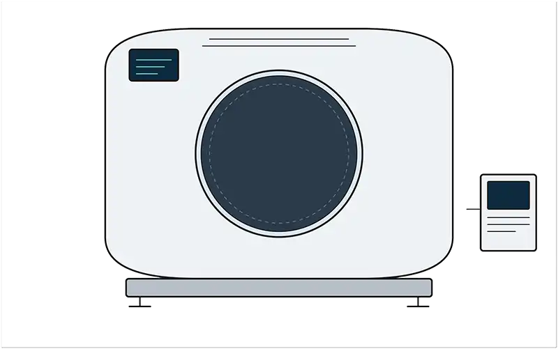
Future developments in diagnostic imaging should be considered in purchasing decisions, but they should not drive them. The key is not to buy the most technologically advanced system available today, but to avoid purchasing equipment that limits how the practice can evolve over the next five to 10 years.
This is particularly relevant in ultrasound. 3D and 4D ultrasound are already well established in human medicine, and veterinary applications are gradually expanding. In dogs, real-time 3D echocardiography has been investigated for cardiac assessments and urinary bladder volume measurements in dogs and cats.
For most first-opinion practices, this does not yet mean that 3D ultrasound should be a purchasing priority. It does, however, mean that practices should ask whether a system is upgradeable.
Can additional probes be added later? Can cardiac software, ECG integration, contrast-enhanced ultrasound or elastography packages be installed if the caseload and expertise develop? Is the machine capable of exporting high-quality clips and still images in standard formats for review, teaching or referral?
Future-proofing ultrasound is less about buying every feature now, and more about avoiding a closed system that cannot adapt to change.
Positron emission tomography (PET-CT) is another example of a modality that may become increasingly common in veterinary medicine, especially in oncology. PET-CT adds functional or metabolic to anatomical information using radiopharmaceutical tracers.
PET-CT has shown promise for improved malignancy detection and more complete staging of canine cancer, although clinical use remains concentrated in specialist and academic environments rather than general practice.
The investment and implications are substantial, and while it is important to understand as an emerging referral pathway, PET-CT is not yet a realistic in-house purchase consideration.
In short, practices should buy equipment with adaptability in mind. A good imaging investment should meet requirements for today’s personnel and patients, and integrate cleanly into existing systems and workflows.
Avoid over-investing in equipment and features that cannot yet be well utilised, but leave room for clinical, technical and technological developments to meet the needs of tomorrow’s clinicians and patients.
Practices should also consider whether future access is better achieved through collaboration rather than ownership. Peripatetic ultrasound, visiting CT or MRI services, referral partnerships, teleradiology, remote scan guidance and shared use of advanced equipment may be alternative solutions. These options can provide clinical benefit without exposing the practice to the full financial and regulatory burden of purchase. This is especially relevant for modalities with high fixed costs and uncertain case volume.
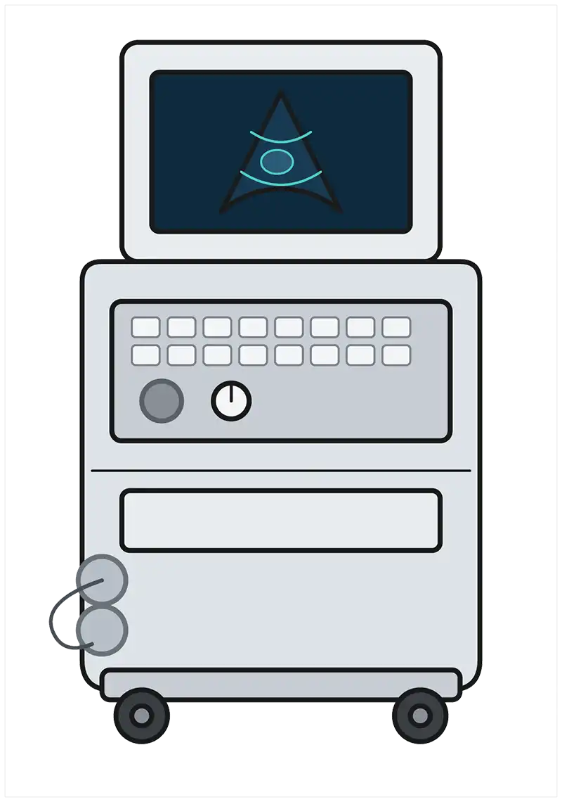
The best investment decision is always guided by robust clinical and financial governance. Analyse your caseload, model the break-even case volume and include the full life cycle cost.
Consider workflows – digital, temporal and physical – staffing, training, safety, reporting, downtime, client affordability and environmental impact. Ask what treatment pathway follows the diagnosis.
The machine is only one component. The true diagnostic tool is a complex system, including trained operators, appropriate case selection, good histories, safe acquisition, expert interpretation and a clear answer to the most important question in imaging – what do we do next?
The right imaging system for the right caseload with the right team can support engaged staff, a high standard of care and improved patient outcomes and client satisfaction, which all contribute to revenue.
Matt Winter is a small animal radiologist with a deep passion for the veterinary profession and veterinary diagnostic imaging in particular. Dr Winter is North American lead and chief veterinary medical officer for VET.CT.